 Movie
Movie Controller

[English] 日本語
 Yorodumi
- PDB-7dam: Adenosine triphosphate phosphoribosyltransferase from Vibrio cholerae -
+ Open data

- Basic information
| Entry | Database: PDB / ID: 7dam | ||||||
|---|---|---|---|---|---|---|---|
| Title | Adenosine triphosphate phosphoribosyltransferase from Vibrio cholerae | ||||||
 Components Components | ATP phosphoribosyltransferase | ||||||
 Keywords Keywords | TRANSFERASE / Adenosine triphosphate phosphoribosyltransferase | ||||||
| Function / homology |  Function and homology information Function and homology informationATP phosphoribosyltransferase / ATP phosphoribosyltransferase activity / L-histidine biosynthetic process / magnesium ion binding / ATP binding / cytoplasm Similarity search - Function | ||||||
| Biological species |  Vibrio cholerae serotype O1 (bacteria) Vibrio cholerae serotype O1 (bacteria) | ||||||
| Method |  X-RAY DIFFRACTION / MOLECULAR REPLACEMENT / molecular replacement / Resolution: 2.7 Å X-RAY DIFFRACTION / MOLECULAR REPLACEMENT / molecular replacement / Resolution: 2.7 Å | ||||||
 Authors Authors | Pal, R.K. / Gourinath, S. / Biswal, B.K. | ||||||
 Citation Citation | Journal: To Be Published Title: Adenosine triphosphate phosphoribosyltransferase from Vibrio cholerae Authors: Pal, R.K. / Gourinath, S. / Biswal, B.K. | ||||||
| History |
|
- Structure visualization
| Structure viewer | Molecule: MolmilJmol/JSmol |
|---|
- Downloads & links
-Download
| PDBx/mmCIF format | 7dam.cif.gz | 112.9 KB | Display | PDBx/mmCIF format |
|---|---|---|---|---|
| PDB format | pdb7dam.ent.gz | 82.2 KB | Display | PDB format |
| PDBx/mmJSON format | 7dam.json.gz | Tree view | PDBx/mmJSON format | |
| Others |  Other downloads Other downloads |
-Validation report
| Arichive directory | https://data.pdbj.org/pub/pdb/validation_reports/da/7damftp://data.pdbj.org/pub/pdb/validation_reports/da/7dam | HTTPS FTP |
|---|
-Related structure data
| Related structure data |  1h3dS S: Starting model for refinement |
|---|---|
| Similar structure data |


-Links
 PDBj
PDBj- Assembly
| Deposited unit | 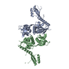
| ||||||||||||||||||
|---|---|---|---|---|---|---|---|---|---|---|---|---|---|---|---|---|---|---|---|
| 1 | 
| ||||||||||||||||||
| Unit cell |
| ||||||||||||||||||
| Noncrystallographic symmetry (NCS) | NCS domain:
NCS domain segments: Component-ID: 1 / Ens-ID: 1 / Beg auth comp-ID: GLN / Beg label comp-ID: GLN / End auth comp-ID: MET / End label comp-ID: MET / Refine code: _ / Auth seq-ID: 10 - 303 / Label seq-ID: 16 - 309
|
-Components
| #1: Protein | Mass: 34579.195 Da / Num. of mol.: 2 Source method: isolated from a genetically manipulated source Source: (gene. exp.) Vibrio cholerae serotype O1 (strain M66-2) (bacteria)Strain: M66-2 / Gene: hisG, VCM66_1088 / Production host: #2: Water | ChemComp-HOH / |  Mass: 18.015 Da / Num. of mol.: 10 / Source method: isolated from a natural source / Formula: H2O Mass: 18.015 Da / Num. of mol.: 10 / Source method: isolated from a natural source / Formula: H2O |
|---|
-Experimental details
-Experiment
| Experiment | Method: X-RAY DIFFRACTION / Number of used crystals: 1 |
|---|
- Sample preparation
| Crystal | Density Matthews: 2.39 Å3/Da / Density % sol: 48.51 % |
|---|---|
| Crystal grow | Temperature: 293.15 K / Method: vapor diffusion Details: 0.1 M Sodium acetate trihydrate pH 4.6 2.0 M Sodium chloride |
-Data collection
| Diffraction | Mean temperature: 100 K / Serial crystal experiment: N | |||||||||||||||
|---|---|---|---|---|---|---|---|---|---|---|---|---|---|---|---|---|
| Diffraction source | Source: ROTATING ANODE / Type: RIGAKU FR-E+ SUPERBRIGHT / Wavelength: 1.54178 Å | |||||||||||||||
| Detector | Type: RIGAKU RAXIS IV++ / Detector: IMAGE PLATE / Date: Nov 3, 2015 | |||||||||||||||
| Radiation | Protocol: SINGLE WAVELENGTH / Monochromatic (M) / Laue (L): M / Scattering type: x-ray | |||||||||||||||
| Radiation wavelength | Wavelength: 1.54178 Å / Relative weight: 1 | |||||||||||||||
| Reflection twin |
| |||||||||||||||
| Reflection | Resolution: 2.7→50 Å / Num. obs: 17005 / % possible obs: 99.7 % / Redundancy: 5.1 % / CC1/2: 0.995 / Net I/σ(I): 14.54 | |||||||||||||||
| Reflection shell | Resolution: 2.7→2.8 Å / Num. unique obs: 1706 / CC1/2: 0.451 |
-Phasing
| Phasing | Method: molecular replacement |
|---|
- Processing
| Software |
| ||||||||||||||||||||||||||||||||||||||||||||||||||||||||||||
|---|---|---|---|---|---|---|---|---|---|---|---|---|---|---|---|---|---|---|---|---|---|---|---|---|---|---|---|---|---|---|---|---|---|---|---|---|---|---|---|---|---|---|---|---|---|---|---|---|---|---|---|---|---|---|---|---|---|---|---|---|---|
| Refinement | Method to determine structure: MOLECULAR REPLACEMENT Starting model: 1H3D Resolution: 2.7→36.67 Å / Cor.coef. Fo:Fc: 0.934 / Cor.coef. Fo:Fc free: 0.903 / SU B: 9.724 / SU ML: 0.219 / Cross valid method: THROUGHOUT / σ(F): 0 / ESU R: 0.534 / ESU R Free: 0.077 / Stereochemistry target values: MAXIMUM LIKELIHOOD Details: HYDROGENS HAVE BEEN ADDED IN THE RIDING POSITIONS U VALUES : REFINED INDIVIDUALLY
| ||||||||||||||||||||||||||||||||||||||||||||||||||||||||||||
| Solvent computation | Ion probe radii: 0.8 Å / Shrinkage radii: 0.8 Å / VDW probe radii: 1.2 Å / Solvent model: MASK | ||||||||||||||||||||||||||||||||||||||||||||||||||||||||||||
| Displacement parameters | Biso max: 134.49 Å2 / Biso mean: 58.298 Å2 / Biso min: 38.28 Å2
| ||||||||||||||||||||||||||||||||||||||||||||||||||||||||||||
| Refinement step | Cycle: final / Resolution: 2.7→36.67 Å
| ||||||||||||||||||||||||||||||||||||||||||||||||||||||||||||
| Refine LS restraints |
| ||||||||||||||||||||||||||||||||||||||||||||||||||||||||||||
| Refine LS restraints NCS | Ens-ID: 1 / Number: 6192 / Refine-ID: X-RAY DIFFRACTION / Type: interatomic distance / Rms dev position: 0.16 Å / Weight position: 0.05
| ||||||||||||||||||||||||||||||||||||||||||||||||||||||||||||
| LS refinement shell | Resolution: 2.705→2.775 Å / Rfactor Rfree error: 0 / Total num. of bins used: 20
|
