[English] 日本語
Yorodumi
- PDB-7d7c: CryoEM structure of gp55-dependent RNA polymerase-promoter open c... -

+
Open data


ID or keywords:

Loading...

-
Basic information

Entry
Database: PDB / ID: 7d7c
TitleCryoEM structure of gp55-dependent RNA polymerase-promoter open complex
Components
  • (DNA-directed RNA polymerase subunit ...Polymerase) x 3
  • DNA (nontemplate strand)
  • DNA (template strand)
  • gp55
KeywordsTRANSCRIPTION / RNA polymerase
Function / homology
Function and homology information


DNA-directed RNA polymerase complex / ribonucleoside binding / DNA-directed 5'-3' RNA polymerase activity / DNA-directed RNA polymerase / protein dimerization activity / DNA-templated transcription / magnesium ion binding / DNA binding / zinc ion binding / cytoplasm
Similarity search - Function
DNA-directed RNA polymerase, subunit beta-prime, bacterial type / DNA-directed RNA polymerase, beta subunit, external 1 domain / RNA polymerase beta subunit external 1 domain / RNA polymerase, alpha subunit, C-terminal / Bacterial RNA polymerase, alpha chain C terminal domain / DNA-directed RNA polymerase, alpha subunit / DNA-directed RNA polymerase beta subunit, bacterial-type / RNA polymerase Rpb1, domain 3 superfamily / RNA polymerase Rpb1, clamp domain superfamily / DNA-directed RNA polymerase, subunit beta-prime ...DNA-directed RNA polymerase, subunit beta-prime, bacterial type / DNA-directed RNA polymerase, beta subunit, external 1 domain / RNA polymerase beta subunit external 1 domain / RNA polymerase, alpha subunit, C-terminal / Bacterial RNA polymerase, alpha chain C terminal domain / DNA-directed RNA polymerase, alpha subunit / DNA-directed RNA polymerase beta subunit, bacterial-type / RNA polymerase Rpb1, domain 3 superfamily / RNA polymerase Rpb1, clamp domain superfamily / DNA-directed RNA polymerase, subunit beta-prime / RNA polymerase Rpb1, domain 3 / RNA polymerase Rpb1, domain 3 / RNA polymerase Rpb2, domain 2 superfamily / RNA polymerase Rpb1, domain 1 / RNA polymerase Rpb1, domain 1 / RNA polymerase, alpha subunit / RNA polymerase Rpb1, domain 4 / RNA polymerase Rpb1, domain 2 / RNA polymerase Rpb1, domain 4 / RNA polymerase, N-terminal / RNA polymerase Rpb1, funnel domain superfamily / RNA polymerase I subunit A N-terminus / RNA polymerase Rpb1, domain 5 / RNA polymerase Rpb1, domain 5 / RNA polymerase, beta subunit, protrusion / RNA polymerase beta subunit / DNA-directed RNA polymerase, insert domain / DNA-directed RNA polymerase, RpoA/D/Rpb3-type / RNA polymerase Rpb3/RpoA insert domain / RNA polymerase Rpb3/Rpb11 dimerisation domain / RNA polymerases D / DNA-directed RNA polymerase, insert domain superfamily / RNA polymerase, RBP11-like subunit / RNA polymerase Rpb2, domain 2 / RNA polymerase Rpb2, domain 2 / RNA polymerase, beta subunit, conserved site / RNA polymerase Rpb2, domain 7 / RNA polymerase Rpb2, domain 3 / RNA polymerase Rpb2, OB-fold / RNA polymerase Rpb2, domain 7 / RNA polymerase Rpb2, domain 3 / RNA polymerases beta chain signature. / DNA-directed RNA polymerase, subunit 2, hybrid-binding domain / DNA-directed RNA polymerase, subunit 2 / DNA-directed RNA polymerase, subunit 2, hybrid-binding domain superfamily / RNA polymerase Rpb2, domain 6
Similarity search - Domain/homology
DNA / DNA (> 10) / DNA-directed RNA polymerase subunit beta / DNA-directed RNA polymerase subunit beta' / DNA-directed RNA polymerase subunit alpha
Similarity search - Component
Biological speciesEscherichia coli (E. coli)
Escherichia coli 1-392-07_S4_C3 (bacteria)
MethodELECTRON MICROSCOPY / single particle reconstruction / cryo EM / Resolution: 3.6 Å
AuthorsShi, J. / Wen, A. / Jin, S. / Feng, Y.
Funding support China, 1items
OrganizationGrant numberCountry
National Natural Science Foundation of China (NSFC)31970040 China
CitationJournal: Nat Commun / Year: 2021
Title: Transcription activation by a sliding clamp.
Authors: Jing Shi / Aijia Wen / Sha Jin / Bo Gao / Yang Huang / Yu Feng /
Abstract: Transcription activation of bacteriophage T4 late genes is accomplished by a transcription activation complex containing RNA polymerase (RNAP), the promoter specificity factor gp55, the coactivator ...Transcription activation of bacteriophage T4 late genes is accomplished by a transcription activation complex containing RNA polymerase (RNAP), the promoter specificity factor gp55, the coactivator gp33, and a universal component of cellular DNA replication, the sliding clamp gp45. Although genetic and biochemical studies have elucidated many aspects of T4 late gene transcription, no precise structure of the transcription machinery in the process is available. Here, we report the cryo-EM structures of a gp55-dependent RNAP-promoter open complex and an intact gp45-dependent transcription activation complex. The structures reveal the interactions between gp55 and the promoter DNA that mediate the recognition of T4 late promoters. In addition to the σR2 homology domain, gp55 has a helix-loop-helix motif that chaperons the template-strand single-stranded DNA of the transcription bubble. Gp33 contacts both RNAP and the upstream double-stranded DNA. Gp45 encircles the DNA and tethers RNAP to it, supporting the idea that gp45 switches the promoter search from three-dimensional diffusion mode to one-dimensional scanning mode.
History
DepositionOct 3, 2020Deposition site: PDBJ / Processing site: PDBJ
Revision 1.0Jan 20, 2021Provider: repository / Type: Initial release
Revision 1.1Mar 17, 2021Group: Database references / Category: citation / citation_author
Item: _citation.journal_volume / _citation.page_first ..._citation.journal_volume / _citation.page_first / _citation.page_last / _citation.pdbx_database_id_DOI / _citation.pdbx_database_id_PubMed / _citation_author.identifier_ORCID
Revision 1.2Mar 27, 2024Group: Data collection / Database references / Category: chem_comp_atom / chem_comp_bond / database_2
Item: _database_2.pdbx_DOI / _database_2.pdbx_database_accession

-
Structure visualization

Movie
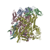
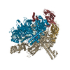
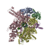
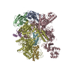
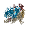
  • Deposited structure unit
  • Imaged by Jmol
  • Download
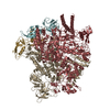
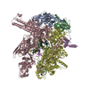
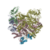
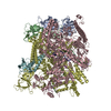
  • Superimposition on EM map
  • EMDB-30604
  • Imaged by UCSF Chimera
  • Download
Movie viewer
Structure viewerMolecule:
MolmilJmol/JSmol

Downloads & links

-
Assembly

Deposited unit
A: DNA-directed RNA polymerase subunit alpha
B: DNA-directed RNA polymerase subunit alpha
C: DNA-directed RNA polymerase subunit beta
D: DNA-directed RNA polymerase subunit beta'
T: DNA (template strand)
F: gp55
N: DNA (nontemplate strand)
hetero molecules


Theoretical massNumber of molelcules
Total (without water)437,38510
Polymers437,2307
Non-polymers1553
Water0
1


  • Idetical with deposited unit
  • defined by author
  • Evidence: native gel electrophoresis
TypeNameSymmetry operationNumber
identity operation1_5551
Buried area37670 Å2
ΔGint-227 kcal/mol
Surface area145140 Å2

-
Components

-
DNA-directed RNA polymerase subunit ... , 3 types, 4 molecules ABCD

#1: Protein DNA-directed RNA polymerase subunit alpha / Polymerase / RNAP subunit alpha / RNA polymerase subunit alpha / Transcriptase subunit alpha


Mass: 36558.680 Da / Num. of mol.: 2
Source method: isolated from a genetically manipulated source
Source: (gene. exp.) Escherichia coli (E. coli) / Production host: Escherichia coli (E. coli) / References: UniProt: U9ZUN7, DNA-directed RNA polymerase
#2: Protein DNA-directed RNA polymerase subunit beta / Polymerase / RNAP subunit beta / RNA polymerase subunit beta / Transcriptase subunit beta


Mass: 150820.875 Da / Num. of mol.: 1
Source method: isolated from a genetically manipulated source
Source: (gene. exp.) Escherichia coli 1-392-07_S4_C3 (bacteria)
Gene: rpoB, AD40_4833 / Production host: Escherichia coli (E. coli)
References: UniProt: A0A080FHH4, DNA-directed RNA polymerase
#3: Protein DNA-directed RNA polymerase subunit beta' / Polymerase / RNAP subunit beta' / RNA polymerase subunit beta' / Transcriptase subunit beta'


Mass: 155366.781 Da / Num. of mol.: 1
Source method: isolated from a genetically manipulated source
Source: (gene. exp.) Escherichia coli (E. coli) / Production host: Escherichia coli (E. coli) / References: UniProt: D7Y6A2, DNA-directed RNA polymerase

-
DNA chain , 2 types, 2 molecules TN

#4: DNA chain DNA (template strand)


Mass: 18203.668 Da / Num. of mol.: 1 / Source method: obtained synthetically / Source: (synth.) Escherichia coli (E. coli)
#6: DNA chain DNA (nontemplate strand)


Mass: 18155.713 Da / Num. of mol.: 1 / Source method: obtained synthetically / Source: (synth.) Escherichia coli (E. coli)

-
Protein , 1 types, 1 molecules F

#5: Protein gp55


Mass: 21565.160 Da / Num. of mol.: 1
Source method: isolated from a genetically manipulated source
Source: (gene. exp.) Escherichia coli (E. coli) / Production host: Escherichia coli (E. coli)

-
Non-polymers , 2 types, 3 molecules

#7: Chemical ChemComp-MG / MAGNESIUM ION


Mass: 24.305 Da / Num. of mol.: 1 / Source method: obtained synthetically / Formula: Mg
#8: Chemical ChemComp-ZN / ZINC ION


Mass: 65.409 Da / Num. of mol.: 2 / Source method: obtained synthetically / Formula: Zn

-
Details

Has ligand of interestN

-
Experimental details

-
Experiment

ExperimentMethod: ELECTRON MICROSCOPY
EM experimentAggregation state: PARTICLE / 3D reconstruction method: single particle reconstruction

-
Sample preparation

ComponentName: gp55-dependent RNA polymerase-promoter open complex / Type: COMPLEX / Entity ID: #1-#6 / Source: MULTIPLE SOURCES
Molecular weightExperimental value: NO
Source (natural)Organism: Thermus thermophilus (bacteria)
Buffer solutionpH: 7.9
SpecimenEmbedding applied: NO / Shadowing applied: NO / Staining applied: NO / Vitrification applied: YES
VitrificationCryogen name: ETHANE

-
Electron microscopy imaging

Experimental equipment
Model: Titan Krios / Image courtesy: FEI Company
MicroscopyModel: FEI TITAN KRIOS
Electron gunElectron source: FIELD EMISSION GUN / Accelerating voltage: 300 kV / Illumination mode: FLOOD BEAM
Electron lensMode: BRIGHT FIELDBright-field microscopy
Image recordingElectron dose: 59 e/Å2 / Film or detector model: GATAN K2 SUMMIT (4k x 4k)

-
Processing

CTF correctionType: PHASE FLIPPING AND AMPLITUDE CORRECTION
3D reconstructionResolution: 3.6 Å / Resolution method: FSC 0.143 CUT-OFF / Num. of particles: 57776 / Symmetry type: POINT

+
About Yorodumi

-
News

-
Feb 9, 2022. New format data for meta-information of EMDB entries

New format data for meta-information of EMDB entries

  • Version 3 of the EMDB header file is now the official format.
  • The previous official version 1.9 will be removed from the archive.

Related info.:EMDB header

External links:wwPDB to switch to version 3 of the EMDB data model

-
Aug 12, 2020. Covid-19 info

Covid-19 info

URL: https://pdbj.org/emnavi/covid19.php

New page: Covid-19 featured information page in EM Navigator.

Related info.:Covid-19 info / Mar 5, 2020. Novel coronavirus structure data

+
Mar 5, 2020. Novel coronavirus structure data

Novel coronavirus structure data

Related info.:Yorodumi Speices / Aug 12, 2020. Covid-19 info

External links:COVID-19 featured content - PDBj / Molecule of the Month (242):Coronavirus Proteases

+
Jan 31, 2019. EMDB accession codes are about to change! (news from PDBe EMDB page)

EMDB accession codes are about to change! (news from PDBe EMDB page)

  • The allocation of 4 digits for EMDB accession codes will soon come to an end. Whilst these codes will remain in use, new EMDB accession codes will include an additional digit and will expand incrementally as the available range of codes is exhausted. The current 4-digit format prefixed with “EMD-” (i.e. EMD-XXXX) will advance to a 5-digit format (i.e. EMD-XXXXX), and so on. It is currently estimated that the 4-digit codes will be depleted around Spring 2019, at which point the 5-digit format will come into force.
  • The EM Navigator/Yorodumi systems omit the EMD- prefix.

Related info.:Q: What is EMD? / ID/Accession-code notation in Yorodumi/EM Navigator

External links:EMDB Accession Codes are Changing Soon! / Contact to PDBj

+
Jul 12, 2017. Major update of PDB

Major update of PDB

  • wwPDB released updated PDB data conforming to the new PDBx/mmCIF dictionary.
  • This is a major update changing the version number from 4 to 5, and with Remediation, in which all the entries are updated.
  • In this update, many items about electron microscopy experimental information are reorganized (e.g. em_software).
  • Now, EM Navigator and Yorodumi are based on the updated data.

External links:wwPDB Remediation / Enriched Model Files Conforming to OneDep Data Standards Now Available in the PDB FTP Archive

-
Yorodumi

Thousand views of thousand structures

  • Yorodumi is a browser for structure data from EMDB, PDB, SASBDB, etc.
  • This page is also the successor to EM Navigator detail page, and also detail information page/front-end page for Omokage search.
  • The word "yorodu" (or yorozu) is an old Japanese word meaning "ten thousand". "mi" (miru) is to see.

Related info.:EMDB / PDB / SASBDB / Comparison of 3 databanks / Yorodumi Search / Aug 31, 2016. New EM Navigator & Yorodumi / Yorodumi Papers / Jmol/JSmol / Function and homology information / Changes in new EM Navigator and Yorodumi

Read more