 Movie
Movie Controller

[English] 日本語
 Yorodumi
- PDB-7d6a: Crystal structure of Oryza sativa Os4BGlu18 monolignol beta-gluco... -
+ Open data

- Basic information
| Entry | Database: PDB / ID: 7d6a | |||||||||
|---|---|---|---|---|---|---|---|---|---|---|
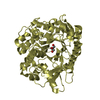
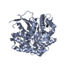
| Title | Crystal structure of Oryza sativa Os4BGlu18 monolignol beta-glucosidase | |||||||||
 Components Components | Beta-glucosidase 18 | |||||||||
 Keywords Keywords | HYDROLASE / monolignol beta-glucosidase / Os4BGlu18 | |||||||||
| Function / homology |  Function and homology information Function and homology informationconiferin metabolic process / coniferin beta-glucosidase activity / glycoside metabolic process / beta-D-fucosidase activity / beta-glucosidase / beta-galactosidase activity / beta-glucosidase activity / carbohydrate metabolic process Similarity search - Function | |||||||||
| Biological species |  | |||||||||
| Method |  X-RAY DIFFRACTION / SYNCHROTRON / MOLECULAR REPLACEMENT / Resolution: 1.7 Å X-RAY DIFFRACTION / SYNCHROTRON / MOLECULAR REPLACEMENT / Resolution: 1.7 Å | |||||||||
 Authors Authors | Baiya, S. / Pengthaisong, S. / Ketudat Cairns, J.R. | |||||||||
| Funding support |  Thailand, 2items Thailand, 2items
| |||||||||
 Citation Citation | Journal: Plos One / Year: 2021 Title: Structural analysis of rice Os4BGlu18 monolignol beta-glucosidase. Authors: Baiya, S. / Pengthaisong, S. / Kitjaruwankul, S. / Ketudat Cairns, J.R. | |||||||||
| History |
|
- Structure visualization
| Structure viewer | Molecule: MolmilJmol/JSmol |
|---|
- Downloads & links
-Download
| PDBx/mmCIF format | 7d6a.cif.gz | 224.7 KB | Display | PDBx/mmCIF format |
|---|---|---|---|---|
| PDB format | pdb7d6a.ent.gz | 175.3 KB | Display | PDB format |
| PDBx/mmJSON format | 7d6a.json.gz | Tree view | PDBx/mmJSON format | |
| Others |  Other downloads Other downloads |
-Validation report
| Arichive directory | https://data.pdbj.org/pub/pdb/validation_reports/d6/7d6aftp://data.pdbj.org/pub/pdb/validation_reports/d6/7d6a | HTTPS FTP |
|---|
-Related structure data
| Related structure data |  7d6bC  2rglS S: Starting model for refinement C: citing same article ( |
|---|---|
| Similar structure data |


-Links
 PDBj
PDBj- Assembly
| Deposited unit | 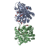
| |||||||||||||||||||||||||||
|---|---|---|---|---|---|---|---|---|---|---|---|---|---|---|---|---|---|---|---|---|---|---|---|---|---|---|---|---|
| 1 | 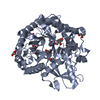
| |||||||||||||||||||||||||||
| 2 | 
| |||||||||||||||||||||||||||
| Unit cell |
| |||||||||||||||||||||||||||
| Noncrystallographic symmetry (NCS) | NCS domain:
NCS domain segments:
|
-Components
| #1: Protein | Mass: 55331.805 Da / Num. of mol.: 2 Source method: isolated from a genetically manipulated source Source: (gene. exp.) Gene: BGLU18, Os04g0513900, LOC_Os04g43410, OSJNBa0004N05.26, OSJNBb0070J16.3 Production host:  #2: Chemical |   Mass: 195.237 Da / Num. of mol.: 2 / Source method: obtained synthetically / Formula: C6H13NO4S / Feature type: SUBJECT OF INVESTIGATION / Comment: pH buffer*YM Mass: 195.237 Da / Num. of mol.: 2 / Source method: obtained synthetically / Formula: C6H13NO4S / Feature type: SUBJECT OF INVESTIGATION / Comment: pH buffer*YM#3: Chemical | ChemComp-GOL /   Mass: 92.094 Da / Num. of mol.: 13 / Source method: obtained synthetically / Formula: C3H8O3 / Feature type: SUBJECT OF INVESTIGATION Mass: 92.094 Da / Num. of mol.: 13 / Source method: obtained synthetically / Formula: C3H8O3 / Feature type: SUBJECT OF INVESTIGATION#4: Chemical | ChemComp-ZN / |   Mass: 65.409 Da / Num. of mol.: 1 / Source method: obtained synthetically / Formula: Zn / Feature type: SUBJECT OF INVESTIGATION Mass: 65.409 Da / Num. of mol.: 1 / Source method: obtained synthetically / Formula: Zn / Feature type: SUBJECT OF INVESTIGATION#5: Water | ChemComp-HOH / |  Mass: 18.015 Da / Num. of mol.: 830 / Source method: isolated from a natural source / Formula: H2O Mass: 18.015 Da / Num. of mol.: 830 / Source method: isolated from a natural source / Formula: H2OHas ligand of interest | Y | Has protein modification | Y | |
|---|
-Experimental details
-Experiment
| Experiment | Method: X-RAY DIFFRACTION / Number of used crystals: 1 |
|---|
- Sample preparation
| Crystal | Density Matthews: 2.05 Å3/Da / Density % sol: 40.09 % |
|---|---|
| Crystal grow | Temperature: 288 K / Method: vapor diffusion, hanging drop / pH: 6 Details: 19% PEG 3350, 0.1M MES, pH 6.0, VAPOR DIFFUSION, HANGING DROP, temperature 288 K |
-Data collection
| Diffraction | Mean temperature: 100 K / Serial crystal experiment: N |
|---|---|
| Diffraction source | Source: SYNCHROTRON / Site: NSRRC  / Beamline: BL15A1 / Wavelength: 1 Å / Beamline: BL15A1 / Wavelength: 1 Å |
| Detector | Type: RAYONIX MX300HE / Detector: CCD / Date: Oct 20, 2016 |
| Radiation | Protocol: SINGLE WAVELENGTH / Monochromatic (M) / Laue (L): M / Scattering type: x-ray |
| Radiation wavelength | Wavelength: 1 Å / Relative weight: 1 |
| Reflection | Resolution: 1.7→30 Å / Num. obs: 96808 / % possible obs: 96 % / Redundancy: 5.4 % / CC1/2: 0.999 / Net I/σ(I): 20.5 |
| Reflection shell | Resolution: 1.7→1.76 Å / Num. unique obs: 9639 / CC1/2: 0.841 |
- Processing
| Software |
| ||||||||||||||||||||||||||||||||||||||||||||||||||||||||||||
|---|---|---|---|---|---|---|---|---|---|---|---|---|---|---|---|---|---|---|---|---|---|---|---|---|---|---|---|---|---|---|---|---|---|---|---|---|---|---|---|---|---|---|---|---|---|---|---|---|---|---|---|---|---|---|---|---|---|---|---|---|---|
| Refinement | Method to determine structure: MOLECULAR REPLACEMENT Starting model: 2RGL Resolution: 1.7→30 Å / Cor.coef. Fo:Fc: 0.97 / Cor.coef. Fo:Fc free: 0.961 / SU B: 1.867 / SU ML: 0.061 / Cross valid method: THROUGHOUT / σ(F): 0 / ESU R: 0.103 / ESU R Free: 0.095 / Stereochemistry target values: MAXIMUM LIKELIHOOD Details: HYDROGENS HAVE BEEN ADDED IN THE RIDING POSITIONS U VALUES : REFINED INDIVIDUALLY
| ||||||||||||||||||||||||||||||||||||||||||||||||||||||||||||
| Solvent computation | Ion probe radii: 0.8 Å / Shrinkage radii: 0.8 Å / VDW probe radii: 1.2 Å / Solvent model: MASK | ||||||||||||||||||||||||||||||||||||||||||||||||||||||||||||
| Displacement parameters | Biso max: 58.2 Å2 / Biso mean: 18.725 Å2 / Biso min: 10.44 Å2
| ||||||||||||||||||||||||||||||||||||||||||||||||||||||||||||
| Refinement step | Cycle: final / Resolution: 1.7→30 Å
| ||||||||||||||||||||||||||||||||||||||||||||||||||||||||||||
| Refine LS restraints |
| ||||||||||||||||||||||||||||||||||||||||||||||||||||||||||||
| Refine LS restraints NCS | Dom-ID: 1 / Auth asym-ID: A / Ens-ID: 1 / Number: 7293 / Refine-ID: X-RAY DIFFRACTION
| ||||||||||||||||||||||||||||||||||||||||||||||||||||||||||||
| LS refinement shell | Resolution: 1.701→1.745 Å / Rfactor Rfree error: 0 / Total num. of bins used: 20
|
