 Movie
Movie Controller

[English] 日本語
 Yorodumi
- PDB-7d5g: Crystal structure of the CsCE with ligand to have a insight into ... -
+ Open data

- Basic information
| Entry | Database: PDB / ID: 7d5g | ||||||
|---|---|---|---|---|---|---|---|
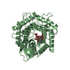
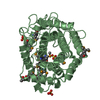
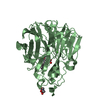
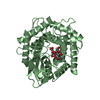
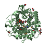
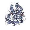
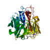
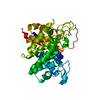
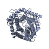
| Title | Crystal structure of the CsCE with ligand to have a insight into the catalytic mechanism | ||||||
 Components Components | Cellobiose 2-epimerase | ||||||
 Keywords Keywords | PROTEIN BINDING / cellobiose 2-epimerase / ligand / epimerization / isomerization | ||||||
| Function / homology | cellobiose epimerase / Cellobiose 2-epimerase / cellobiose epimerase activity / N-acylglucosamine 2-epimerase/Cellobiose 2-epimerase / N-acylglucosamine 2-epimerase (GlcNAc 2-epimerase) / Six-hairpin glycosidase-like superfamily / Six-hairpin glycosidase superfamily / carbohydrate metabolic process / Cellobiose 2-epimerase Function and homology information Function and homology information | ||||||
| Biological species |   Caldicellulosiruptor saccharolyticus DSM 8903 (bacteria) Caldicellulosiruptor saccharolyticus DSM 8903 (bacteria) | ||||||
| Method |  X-RAY DIFFRACTION / SYNCHROTRON / MOLECULAR REPLACEMENT / Resolution: 1.6 Å X-RAY DIFFRACTION / SYNCHROTRON / MOLECULAR REPLACEMENT / Resolution: 1.6 Å | ||||||
 Authors Authors | Yang, R.J. / Feng, Y.H. / Andrew, J.F. | ||||||
| Funding support |  China, 1items China, 1items
| ||||||
 Citation Citation | Journal: Int.J.Biol.Macromol. / Year: 2023 Title: A precise swaying map for how promiscuous cellobiose-2-epimerase operate bi-reaction Authors: Feng, Y. / Lyu, X. / Cong, Y. / Miao, T. / Fang, B. / Zhang, C. / Shen, Q. / Matthews, M. / Fisher, A.J. / Zhang, J.Z. / Zhang, L. / Yang, R. | ||||||
| History |
|
- Structure visualization
| Structure viewer | Molecule: MolmilJmol/JSmol |
|---|
- Downloads & links
-Download
| PDBx/mmCIF format | 7d5g.cif.gz | 108.4 KB | Display | PDBx/mmCIF format |
|---|---|---|---|---|
| PDB format | pdb7d5g.ent.gz | 81.3 KB | Display | PDB format |
| PDBx/mmJSON format | 7d5g.json.gz | Tree view | PDBx/mmJSON format | |
| Others |  Other downloads Other downloads |
-Validation report
| Arichive directory | https://data.pdbj.org/pub/pdb/validation_reports/d5/7d5gftp://data.pdbj.org/pub/pdb/validation_reports/d5/7d5g | HTTPS FTP |
|---|
-Related structure data
| Related structure data | 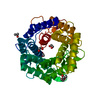 4z4jS S: Starting model for refinement |
|---|---|
| Similar structure data |
-Links
 PDBj
PDBj
- Assembly
| Deposited unit | 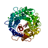
| |||||||||
|---|---|---|---|---|---|---|---|---|---|---|
| 1 |
| |||||||||
| Unit cell |
| |||||||||
| Components on special symmetry positions |
|
-Components
| #1: Protein | Mass: 46469.965 Da / Num. of mol.: 1 Source method: isolated from a genetically manipulated source Source: (gene. exp.) Caldicellulosiruptor saccharolyticus DSM 8903 (bacteria)Strain: DSM 8903 / Gene: Csac_0294 Production host: References: UniProt: A4XGA6, cellobiose epimerase | ||||||
|---|---|---|---|---|---|---|---|
| #2: Polysaccharide | beta-D-glucopyranose-(1-4)-beta-D-fructofuranose Type: oligosaccharide / Mass: 342.297 Da / Num. of mol.: 1 Source method: isolated from a genetically manipulated source | ||||||
| #3: Chemical |   Mass: 35.453 Da / Num. of mol.: 3 / Source method: obtained synthetically / Formula: Cl / Feature type: SUBJECT OF INVESTIGATION Mass: 35.453 Da / Num. of mol.: 3 / Source method: obtained synthetically / Formula: Cl / Feature type: SUBJECT OF INVESTIGATION#4: Water | ChemComp-HOH / |  Mass: 18.015 Da / Num. of mol.: 323 / Source method: isolated from a natural source / Formula: H2O Mass: 18.015 Da / Num. of mol.: 323 / Source method: isolated from a natural source / Formula: H2OHas ligand of interest | Y | Has protein modification | N | |
-Experimental details
-Experiment
| Experiment | Method: X-RAY DIFFRACTION / Number of used crystals: 1 |
|---|
- Sample preparation
| Crystal | Density Matthews: 2.16 Å3/Da / Density % sol: 43.16 % Description: THE ENTRY CONTAINS FRIEDEL PAIRS IN I/F_PLUS/MINUS COLUMNS. |
|---|---|
| Crystal grow | Temperature: 298 K / Method: evaporation / pH: 6.5 / Details: Mgnesium chloride, Bis-HCl, PEG4000 |
-Data collection
| Diffraction | Mean temperature: 273 K / Serial crystal experiment: N |
|---|---|
| Diffraction source | Source: SYNCHROTRON / Site: APS  / Beamline: 24-ID-C / Wavelength: 0.9792 Å / Beamline: 24-ID-C / Wavelength: 0.9792 Å |
| Detector | Type: MARMOSAIC 300 mm CCD / Detector: CCD / Date: Jul 11, 2017 |
| Radiation | Protocol: SINGLE WAVELENGTH / Monochromatic (M) / Laue (L): M / Scattering type: x-ray |
| Radiation wavelength | Wavelength: 0.9792 Å / Relative weight: 1 |
| Reflection | Resolution: 1.6→66.49 Å / Num. obs: 54194 / % possible obs: 99.9 % / Redundancy: 6.48 % / Biso Wilson estimate: 18.73 Å2 / Rmerge(I) obs: 0.092 / Net I/σ(I): 12.2 |
| Reflection shell | Resolution: 1.6→1.63 Å / Redundancy: 6.3 % / Rmerge(I) obs: 1.182 / Mean I/σ(I) obs: 1.8 / Num. unique obs: 2756 / % possible all: 100 |
- Processing
| Software |
| |||||||||||||||||||||||||||||||||||||||||||||||||||||||||||||||||||||||||||||||||||||||||||||||||||||||||||||||||||||||||||||||||||||||||||||||||||
|---|---|---|---|---|---|---|---|---|---|---|---|---|---|---|---|---|---|---|---|---|---|---|---|---|---|---|---|---|---|---|---|---|---|---|---|---|---|---|---|---|---|---|---|---|---|---|---|---|---|---|---|---|---|---|---|---|---|---|---|---|---|---|---|---|---|---|---|---|---|---|---|---|---|---|---|---|---|---|---|---|---|---|---|---|---|---|---|---|---|---|---|---|---|---|---|---|---|---|---|---|---|---|---|---|---|---|---|---|---|---|---|---|---|---|---|---|---|---|---|---|---|---|---|---|---|---|---|---|---|---|---|---|---|---|---|---|---|---|---|---|---|---|---|---|---|---|---|---|
| Refinement | Method to determine structure: MOLECULAR REPLACEMENT Starting model: 4Z4J Resolution: 1.6→66.49 Å / SU ML: 0.15 / Cross valid method: THROUGHOUT / σ(F): 1.35 / Phase error: 18.87 / Stereochemistry target values: ML
| |||||||||||||||||||||||||||||||||||||||||||||||||||||||||||||||||||||||||||||||||||||||||||||||||||||||||||||||||||||||||||||||||||||||||||||||||||
| Solvent computation | Shrinkage radii: 0.9 Å / VDW probe radii: 1.11 Å / Solvent model: FLAT BULK SOLVENT MODEL | |||||||||||||||||||||||||||||||||||||||||||||||||||||||||||||||||||||||||||||||||||||||||||||||||||||||||||||||||||||||||||||||||||||||||||||||||||
| Displacement parameters | Biso max: 90.48 Å2 / Biso mean: 23.5491 Å2 / Biso min: 9.07 Å2 | |||||||||||||||||||||||||||||||||||||||||||||||||||||||||||||||||||||||||||||||||||||||||||||||||||||||||||||||||||||||||||||||||||||||||||||||||||
| Refinement step | Cycle: final / Resolution: 1.6→66.49 Å
| |||||||||||||||||||||||||||||||||||||||||||||||||||||||||||||||||||||||||||||||||||||||||||||||||||||||||||||||||||||||||||||||||||||||||||||||||||
| LS refinement shell | Refine-ID: X-RAY DIFFRACTION / Rfactor Rfree error: 0 / Total num. of bins used: 20
|
