 Movie
Movie Controller


+ Open data

- Basic information
| Entry | Database: PDB / ID: 7d3y | ||||||
|---|---|---|---|---|---|---|---|
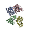
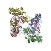
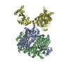
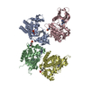
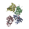
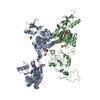
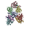
| Title | Crystal structure of the osPHR2-osSPX2 complex | ||||||
 Components Components |
| ||||||
 Keywords Keywords | DNA BINDING PROTEIN / Protein PHOSPHATE STARVATION RESPONSE 2 / MYB / SPX2 | ||||||
| Function / homology |  Function and homology information Function and homology informationregulation of leaf development / negative regulation of unidimensional cell growth / response to nitrate / regulation of phosphate transport / cellular response to phosphate starvation / cellular response to cold / DNA-binding transcription factor activity / DNA binding / nucleus / cytoplasm Similarity search - Function | ||||||
| Biological species |   Homo sapiens (human) Homo sapiens (human) | ||||||
| Method |  X-RAY DIFFRACTION / SYNCHROTRON / MOLECULAR REPLACEMENT / Resolution: 3.11 Å X-RAY DIFFRACTION / SYNCHROTRON / MOLECULAR REPLACEMENT / Resolution: 3.11 Å | ||||||
 Authors Authors | Zhang, Q.X. / Guan, Z.Y. / Zuo, J.Q. / Zhang, Z.F. / Liu, Z. | ||||||
 Citation Citation | Journal: Nat Commun / Year: 2022 Title: Mechanistic insights into the regulation of plant phosphate homeostasis by the rice SPX2 - PHR2 complex. Authors: Guan, Z. / Zhang, Q. / Zhang, Z. / Zuo, J. / Chen, J. / Liu, R. / Savarin, J. / Broger, L. / Cheng, P. / Wang, Q. / Pei, K. / Zhang, D. / Zou, T. / Yan, J. / Yin, P. / Hothorn, M. / Liu, Z. | ||||||
| History |
|
- Structure visualization
| Structure viewer | Molecule: MolmilJmol/JSmol |
|---|
- Downloads & links
-Download
| PDBx/mmCIF format | 7d3y.cif.gz | 460.9 KB | Display | PDBx/mmCIF format |
|---|---|---|---|---|
| PDB format | pdb7d3y.ent.gz | 322.2 KB | Display | PDB format |
| PDBx/mmJSON format | 7d3y.json.gz | Tree view | PDBx/mmJSON format | |
| Others |  Other downloads Other downloads |
-Validation report
| Summary document | 7d3y_validation.pdf.gz | 1.2 MB | Display | wwPDB validaton report |
|---|---|---|---|---|
| Full document | 7d3y_full_validation.pdf.gz | 1.2 MB | Display | |
| Data in XML | 7d3y_validation.xml.gz | 31.3 KB | Display | |
| Data in CIF | 7d3y_validation.cif.gz | 42.5 KB | Display | |
| Arichive directory | https://data.pdbj.org/pub/pdb/validation_reports/d3/7d3yftp://data.pdbj.org/pub/pdb/validation_reports/d3/7d3y | HTTPS FTP |
-Related structure data
| Related structure data | 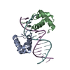 7d3tSC S: Starting model for refinement C: citing same article ( |
|---|---|
| Similar structure data |
-Links
 PDBj
PDBj


- Assembly
| Deposited unit | 
| ||||||||||||
|---|---|---|---|---|---|---|---|---|---|---|---|---|---|
| 1 |
| ||||||||||||
| Unit cell |
|
-Components
| #1: Protein | Mass: 43437.926 Da / Num. of mol.: 2 / Fragment: macro domain Source method: isolated from a genetically manipulated source Details: Fusion protein of SPX2, linker and histone macroH2A1.1 Source: (gene. exp.) Homo sapiens (human)Gene: SPX2, OsI_06282, MACROH2A1, H2AFY / Production host:  #2: Protein | Mass: 16809.039 Da / Num. of mol.: 3 Source method: isolated from a genetically manipulated source Source: (gene. exp.) Gene: PHR2, Os07g0438800, LOC_Os07g25710, OSJNBa0026I22.19, P0443H10.4 Production host: #3: Chemical |   Mass: 660.035 Da / Num. of mol.: 2 / Source method: obtained synthetically / Formula: C6H18O24P6 Mass: 660.035 Da / Num. of mol.: 2 / Source method: obtained synthetically / Formula: C6H18O24P6 |
|---|
-Experimental details
-Experiment
| Experiment | Method: X-RAY DIFFRACTION / Number of used crystals: 1 |
|---|
- Sample preparation
| Crystal | Density Matthews: 3.29 Å3/Da / Density % sol: 62.63 % |
|---|---|
| Crystal grow | Temperature: 291.15 K / Method: vapor diffusion, hanging drop Details: Ammonium sulfate, Pentaerythritol ethoxylate, Tacsimate PH range: 7 |
-Data collection
| Diffraction | Mean temperature: 100 K / Serial crystal experiment: N |
|---|---|
| Diffraction source | Source: SYNCHROTRON / Site: SSRF  / Beamline: BL17U1 / Wavelength: 0.9792 Å / Beamline: BL17U1 / Wavelength: 0.9792 Å |
| Detector | Type: DECTRIS EIGER X 16M / Detector: PIXEL / Date: Mar 18, 2019 / Details: 0.9792 |
| Radiation | Protocol: SINGLE WAVELENGTH / Monochromatic (M) / Laue (L): M / Scattering type: x-ray |
| Radiation wavelength | Wavelength: 0.9792 Å / Relative weight: 1 |
| Reflection | Resolution: 3.11→48.38 Å / Num. obs: 32993 / % possible obs: 99.9 % / Redundancy: 19.8 % / Biso Wilson estimate: 123.72 Å2 / CC1/2: 0.999 / Rmerge(I) obs: 0.049 / Rpim(I) all: 0.012 / Rrim(I) all: 0.051 / Net I/σ(I): 35.4 |
| Reflection shell | Resolution: 3.11→3.28 Å / Rmerge(I) obs: 0.844 / Mean I/σ(I) obs: 4.7 / Num. unique obs: 4743 / CC1/2: 0.957 / Rpim(I) all: 0.188 / Rrim(I) all: 0.865 |
- Processing
| Software |
| |||||||||||||||||||||||||||||||||||||||||||||||||||||||||||||||||||||||||||||||||||||||||||
|---|---|---|---|---|---|---|---|---|---|---|---|---|---|---|---|---|---|---|---|---|---|---|---|---|---|---|---|---|---|---|---|---|---|---|---|---|---|---|---|---|---|---|---|---|---|---|---|---|---|---|---|---|---|---|---|---|---|---|---|---|---|---|---|---|---|---|---|---|---|---|---|---|---|---|---|---|---|---|---|---|---|---|---|---|---|---|---|---|---|---|---|---|
| Refinement | Method to determine structure: MOLECULAR REPLACEMENT Starting model: 7D3T Resolution: 3.11→48.38 Å / SU ML: 0.4359 / Cross valid method: FREE R-VALUE / σ(F): 1.34 / Phase error: 33.1497 Stereochemistry target values: GeoStd + Monomer Library + CDL v1.2
| |||||||||||||||||||||||||||||||||||||||||||||||||||||||||||||||||||||||||||||||||||||||||||
| Solvent computation | Shrinkage radii: 0.9 Å / VDW probe radii: 1.11 Å / Solvent model: FLAT BULK SOLVENT MODEL | |||||||||||||||||||||||||||||||||||||||||||||||||||||||||||||||||||||||||||||||||||||||||||
| Displacement parameters | Biso mean: 147.49 Å2 | |||||||||||||||||||||||||||||||||||||||||||||||||||||||||||||||||||||||||||||||||||||||||||
| Refinement step | Cycle: LAST / Resolution: 3.11→48.38 Å
| |||||||||||||||||||||||||||||||||||||||||||||||||||||||||||||||||||||||||||||||||||||||||||
| Refine LS restraints |
| |||||||||||||||||||||||||||||||||||||||||||||||||||||||||||||||||||||||||||||||||||||||||||
| LS refinement shell |
| |||||||||||||||||||||||||||||||||||||||||||||||||||||||||||||||||||||||||||||||||||||||||||
| Refinement TLS params. | Method: refined / Origin x: 26.7645188018 Å / Origin y: -60.0185349528 Å / Origin z: 24.513360751 Å
| |||||||||||||||||||||||||||||||||||||||||||||||||||||||||||||||||||||||||||||||||||||||||||
| Refinement TLS group | Selection details: all |
