 Movie
Movie Controller


+ Open data

- Basic information
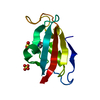
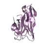
| Entry | Database: PDB / ID: 7d2a | ||||||
|---|---|---|---|---|---|---|---|
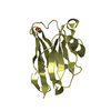
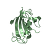
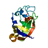
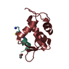
| Title | CBM32 of AlyQ in complex with 4,5-unsaturated mannuronic acid | ||||||
 Components Components | AlyQ | ||||||
 Keywords Keywords | SUGAR BINDING PROTEIN / CBM32 / alginate | ||||||
| Function / homology |  Function and homology information Function and homology informationhydrolase activity, hydrolyzing O-glycosyl compounds / carbohydrate metabolic process / metal ion binding Similarity search - Function | ||||||
| Biological species |  Persicobacter sp. CCB-QB2 (bacteria) Persicobacter sp. CCB-QB2 (bacteria) | ||||||
| Method |  X-RAY DIFFRACTION / MOLECULAR REPLACEMENT / Resolution: 1.57 Å X-RAY DIFFRACTION / MOLECULAR REPLACEMENT / Resolution: 1.57 Å | ||||||
 Authors Authors | Teh, A.H. / Sim, P.F. | ||||||
| Funding support |  Malaysia, 1items Malaysia, 1items
| ||||||
 Citation Citation | Journal: Biochem.Biophys.Res.Commun. / Year: 2020 Title: Structural basis for binding uronic acids by family 32 carbohydrate-binding modules. Authors: Teh, A.H. / Sim, P.F. / Hisano, T. | ||||||
| History |
|
- Structure visualization
| Structure viewer | Molecule: MolmilJmol/JSmol |
|---|
- Downloads & links
-Download
| PDBx/mmCIF format | 7d2a.cif.gz | 47.7 KB | Display | PDBx/mmCIF format |
|---|---|---|---|---|
| PDB format | pdb7d2a.ent.gz | 29.3 KB | Display | PDB format |
| PDBx/mmJSON format | 7d2a.json.gz | Tree view | PDBx/mmJSON format | |
| Others |  Other downloads Other downloads |
-Validation report
| Summary document | 7d2a_validation.pdf.gz | 784.6 KB | Display | wwPDB validaton report |
|---|---|---|---|---|
| Full document | 7d2a_full_validation.pdf.gz | 784.9 KB | Display | |
| Data in XML | 7d2a_validation.xml.gz | 9.4 KB | Display | |
| Data in CIF | 7d2a_validation.cif.gz | 13.2 KB | Display | |
| Arichive directory | https://data.pdbj.org/pub/pdb/validation_reports/d2/7d2aftp://data.pdbj.org/pub/pdb/validation_reports/d2/7d2a | HTTPS FTP |
-Related structure data
| Related structure data |  7d29C  5xnrS S: Starting model for refinement C: citing same article ( |
|---|---|
| Similar structure data |


-Links
 PDBj
PDBj

- Assembly
| Deposited unit | 
| ||||||||
|---|---|---|---|---|---|---|---|---|---|
| 1 |
| ||||||||
| Unit cell |
|
-Components
-Protein / Sugars , 2 types, 2 molecules A
| #1: Protein | Mass: 16095.180 Da / Num. of mol.: 1 / Fragment: CBM32 Source method: isolated from a genetically manipulated source Source: (gene. exp.) Persicobacter sp. CCB-QB2 (bacteria) / Production host: |
|---|---|
| #2: Polysaccharide | 4-deoxy-alpha-L-erythro-hex-4-enopyranuronic acid-(1-4)-alpha-L-gulopyranuronic acid Source method: isolated from a genetically manipulated source |
-Non-polymers , 4 types, 163 molecules 


| #3: Chemical | ChemComp-CA /  Mass: 40.078 Da / Num. of mol.: 1 / Source method: obtained synthetically / Formula: Ca Mass: 40.078 Da / Num. of mol.: 1 / Source method: obtained synthetically / Formula: Ca |
|---|---|
| #4: Chemical | ChemComp-PG4 /  Mass: 194.226 Da / Num. of mol.: 1 / Source method: obtained synthetically / Formula: C8H18O5 / Comment: precipitant*YM Mass: 194.226 Da / Num. of mol.: 1 / Source method: obtained synthetically / Formula: C8H18O5 / Comment: precipitant*YM |
| #5: Chemical | ChemComp-NA /  Mass: 22.990 Da / Num. of mol.: 1 / Source method: obtained synthetically / Formula: Na Mass: 22.990 Da / Num. of mol.: 1 / Source method: obtained synthetically / Formula: Na |
| #6: Water | ChemComp-HOH / Mass: 18.015 Da / Num. of mol.: 160 / Source method: isolated from a natural source / Formula: H2O |
-Details
| Has ligand of interest | Y |
|---|
-Experimental details
-Experiment
| Experiment | Method: X-RAY DIFFRACTION / Number of used crystals: 1 |
|---|
- Sample preparation
| Crystal | Density Matthews: 2.21 Å3/Da / Density % sol: 44.27 % |
|---|---|
| Crystal grow | Temperature: 293 K / Method: vapor diffusion, sitting drop / pH: 7.5 Details: 0.1M sodium HEPES, 2% PEG 400, 2.0M ammonium sulphate, 10mg/ml enzymatically degraded sodium alginate |
-Data collection
| Diffraction | Mean temperature: 100 K / Serial crystal experiment: N |
|---|---|
| Diffraction source | Source: ROTATING ANODE / Type: RIGAKU MICROMAX-007 HF / Wavelength: 1.54 Å |
| Detector | Type: RIGAKU RAXIS IV++ / Detector: IMAGE PLATE / Date: May 30, 2017 |
| Radiation | Protocol: SINGLE WAVELENGTH / Monochromatic (M) / Laue (L): M / Scattering type: x-ray |
| Radiation wavelength | Wavelength: 1.54 Å / Relative weight: 1 |
| Reflection | Resolution: 1.57→40.34 Å / Num. obs: 18779 / % possible obs: 91.5 % / Redundancy: 6.73 % / Rmerge(I) obs: 0.043 / Rrim(I) all: 0.046 / Χ2: 0.95 / Net I/σ(I): 22.2 |
| Reflection shell | Resolution: 1.57→1.63 Å / Redundancy: 5.97 % / Rmerge(I) obs: 0.203 / Mean I/σ(I) obs: 6.9 / Num. unique obs: 1284 / Rrim(I) all: 0.223 / Χ2: 1.19 / % possible all: 63.6 |
- Processing
| Software |
| ||||||||||||||||||||
|---|---|---|---|---|---|---|---|---|---|---|---|---|---|---|---|---|---|---|---|---|---|
| Refinement | Method to determine structure: MOLECULAR REPLACEMENT Starting model: 5XNR Resolution: 1.57→40.34 Å / Cor.coef. Fo:Fc: 0.961 / Cor.coef. Fo:Fc free: 0.949 / SU B: 1.107 / SU ML: 0.041 / Cross valid method: FREE R-VALUE / ESU R: 0.084 / ESU R Free: 0.083
| ||||||||||||||||||||
| Solvent computation | Ion probe radii: 0.8 Å / Shrinkage radii: 0.8 Å / VDW probe radii: 1.2 Å / Solvent model: MASK BULK SOLVENT | ||||||||||||||||||||
| Displacement parameters | Biso mean: 14.486 Å2
| ||||||||||||||||||||
| Refinement step | Cycle: LAST / Resolution: 1.57→40.34 Å
| ||||||||||||||||||||
| LS refinement shell | Resolution: 1.57→1.611 Å
|
