 Movie
Movie Controller


+ Open data

- Basic information
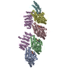
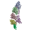
| Entry | Database: PDB / ID: 7cym | ||||||
|---|---|---|---|---|---|---|---|
| Title | Crystal structure of LI-Cadherin EC1-4 | ||||||
 Components Components | Cadherin-17 | ||||||
 Keywords Keywords | CELL ADHESION / LI-cadherin / dimerization / MD simulations / SAXS / cell-adhesion / analytical ultracentrifugation / protein chemistry | ||||||
| Function / homology |  Function and homology information Function and homology informationoligopeptide transmembrane transport / positive regulation of integrin activation by cell surface receptor linked signal transduction / proton-dependent oligopeptide secondary active transmembrane transporter activity / marginal zone B cell differentiation / germinal center B cell differentiation / calcium-dependent cell-cell adhesion / cell-cell adhesion mediated by cadherin / adherens junction organization / catenin complex / cell-cell junction assembly ...oligopeptide transmembrane transport / positive regulation of integrin activation by cell surface receptor linked signal transduction / proton-dependent oligopeptide secondary active transmembrane transporter activity / marginal zone B cell differentiation / germinal center B cell differentiation / calcium-dependent cell-cell adhesion / cell-cell adhesion mediated by cadherin / adherens junction organization / catenin complex / cell-cell junction assembly / Adherens junctions interactions / homophilic cell-cell adhesion / spleen development / integrin-mediated signaling pathway / adherens junction / beta-catenin binding / integrin binding / cell morphogenesis / cell junction / cell migration / basolateral plasma membrane / cell adhesion / cadherin binding / calcium ion binding / cell surface / nucleoplasm / plasma membrane Similarity search - Function | ||||||
| Biological species |  Homo sapiens (human) Homo sapiens (human) | ||||||
| Method |  X-RAY DIFFRACTION / SYNCHROTRON / MOLECULAR REPLACEMENT / Resolution: 2.7 Å X-RAY DIFFRACTION / SYNCHROTRON / MOLECULAR REPLACEMENT / Resolution: 2.7 Å | ||||||
 Authors Authors | Caaveiro, J.M.M. / Yui, A. / Tsumoto, K. | ||||||
 Citation Citation | Journal: J.Biol.Chem. / Year: 2021 Title: Mechanism of dimerization and structural features of human LI-cadherin. Authors: Yui, A. / Caaveiro, J.M.M. / Kuroda, D. / Nakakido, M. / Nagatoishi, S. / Goda, S. / Maruno, T. / Uchiyama, S. / Tsumoto, K. | ||||||
| History |
|
- Structure visualization
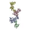
| Structure viewer | Molecule: MolmilJmol/JSmol |
|---|
- Downloads & links
-Download
| PDBx/mmCIF format | 7cym.cif.gz | 357.8 KB | Display | PDBx/mmCIF format |
|---|---|---|---|---|
| PDB format | pdb7cym.ent.gz | 294.1 KB | Display | PDB format |
| PDBx/mmJSON format | 7cym.json.gz | Tree view | PDBx/mmJSON format | |
| Others |  Other downloads Other downloads |
-Validation report
| Summary document | 7cym_validation.pdf.gz | 4.5 MB | Display | wwPDB validaton report |
|---|---|---|---|---|
| Full document | 7cym_full_validation.pdf.gz | 4.6 MB | Display | |
| Data in XML | 7cym_validation.xml.gz | 33.2 KB | Display | |
| Data in CIF | 7cym_validation.cif.gz | 45.3 KB | Display | |
| Arichive directory | https://data.pdbj.org/pub/pdb/validation_reports/cy/7cymftp://data.pdbj.org/pub/pdb/validation_reports/cy/7cym | HTTPS FTP |
-Related structure data
| Related structure data |  7ev1C  4zmyS S: Starting model for refinement C: citing same article ( |
|---|---|
| Similar structure data |
-Links
 PDBj
PDBj

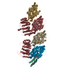
- Assembly
| Deposited unit | 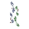
| |||||||||||||||||||||||||||
|---|---|---|---|---|---|---|---|---|---|---|---|---|---|---|---|---|---|---|---|---|---|---|---|---|---|---|---|---|
| 1 |
| |||||||||||||||||||||||||||
| Unit cell |
| |||||||||||||||||||||||||||
| Noncrystallographic symmetry (NCS) | NCS domain:
NCS domain segments:
|
-Components
-Protein , 1 types, 2 molecules AB
| #1: Protein | Mass: 49874.664 Da / Num. of mol.: 2 Source method: isolated from a genetically manipulated source Source: (gene. exp.) Homo sapiens (human) / Gene: CDH17 / Cell line (production host): EXPI293 / Production host: Homo sapiens (human) / References: UniProt: Q12864 |
|---|
-Sugars , 3 types, 6 molecules 
| #2: Polysaccharide | Source method: isolated from a genetically manipulated source #3: Polysaccharide | 2-acetamido-2-deoxy-beta-D-glucopyranose-(1-4)-2-acetamido-2-deoxy-beta-D-glucopyranose | Source method: isolated from a genetically manipulated source #4: Sugar |  Type: D-saccharide, beta linking / Mass: 221.208 Da / Num. of mol.: 3 / Source method: obtained synthetically / Formula: C8H15NO6 Type: D-saccharide, beta linking / Mass: 221.208 Da / Num. of mol.: 3 / Source method: obtained synthetically / Formula: C8H15NO6 |
|---|
-Non-polymers , 2 types, 45 molecules 

| #5: Chemical | ChemComp-CA /  Mass: 40.078 Da / Num. of mol.: 12 / Source method: obtained synthetically / Formula: Ca / Feature type: SUBJECT OF INVESTIGATION Mass: 40.078 Da / Num. of mol.: 12 / Source method: obtained synthetically / Formula: Ca / Feature type: SUBJECT OF INVESTIGATION#6: Water | ChemComp-HOH / | Mass: 18.015 Da / Num. of mol.: 33 / Source method: isolated from a natural source / Formula: H2O |
|---|
-Details
| Has ligand of interest | Y |
|---|---|
| Has protein modification | Y |
-Experimental details
-Experiment
| Experiment | Method: X-RAY DIFFRACTION / Number of used crystals: 1 |
|---|
- Sample preparation
| Crystal | Density Matthews: 3.79 Å3/Da / Density % sol: 67.51 % |
|---|---|
| Crystal grow | Temperature: 293.15 K / Method: vapor diffusion, hanging drop / pH: 7.5 Details: 200 mM sodium sulfate, 20% w/v Polyethylene glycol 3350 |
-Data collection
| Diffraction | Mean temperature: 100 K / Serial crystal experiment: N |
|---|---|
| Diffraction source | Source: SYNCHROTRON / Site: Photon Factory  / Beamline: BL-5A / Wavelength: 1 Å / Beamline: BL-5A / Wavelength: 1 Å |
| Detector | Type: DECTRIS PILATUS3 6M / Detector: PIXEL / Date: Jun 23, 2018 |
| Radiation | Protocol: SINGLE WAVELENGTH / Monochromatic (M) / Laue (L): M / Scattering type: x-ray |
| Radiation wavelength | Wavelength: 1 Å / Relative weight: 1 |
| Reflection | Resolution: 2.7→59.2 Å / Num. obs: 41272 / % possible obs: 99.9 % / Redundancy: 6.1 % / CC1/2: 0.998 / Rmerge(I) obs: 0.095 / Rpim(I) all: 0.041 / Net I/σ(I): 1.8 |
| Reflection shell | Resolution: 2.7→2.85 Å / Redundancy: 6.2 % / Rmerge(I) obs: 0.858 / Mean I/σ(I) obs: 1.8 / Num. unique obs: 5955 / CC1/2: 0.907 / Rpim(I) all: 0.371 / % possible all: 100 |
- Processing
| Software |
| |||||||||||||||||||||||||||||||||||||||||||||||||||||||||||||||||||||||||||
|---|---|---|---|---|---|---|---|---|---|---|---|---|---|---|---|---|---|---|---|---|---|---|---|---|---|---|---|---|---|---|---|---|---|---|---|---|---|---|---|---|---|---|---|---|---|---|---|---|---|---|---|---|---|---|---|---|---|---|---|---|---|---|---|---|---|---|---|---|---|---|---|---|---|---|---|---|
| Refinement | Method to determine structure: MOLECULAR REPLACEMENT Starting model: 4ZMY Resolution: 2.7→55.25 Å / Cor.coef. Fo:Fc: 0.934 / Cor.coef. Fo:Fc free: 0.891 / SU B: 35.033 / SU ML: 0.311 / Cross valid method: THROUGHOUT / σ(F): 0 / ESU R: 0.468 / ESU R Free: 0.316 / Stereochemistry target values: MAXIMUM LIKELIHOOD Details: HYDROGENS HAVE BEEN ADDED IN THE RIDING POSITIONS U VALUES : WITH TLS ADDED
| |||||||||||||||||||||||||||||||||||||||||||||||||||||||||||||||||||||||||||
| Solvent computation | Ion probe radii: 0.8 Å / Shrinkage radii: 0.8 Å / VDW probe radii: 1.2 Å / Solvent model: MASK | |||||||||||||||||||||||||||||||||||||||||||||||||||||||||||||||||||||||||||
| Displacement parameters | Biso max: 146.91 Å2 / Biso mean: 75.276 Å2 / Biso min: 44.55 Å2
| |||||||||||||||||||||||||||||||||||||||||||||||||||||||||||||||||||||||||||
| Refinement step | Cycle: final / Resolution: 2.7→55.25 Å
| |||||||||||||||||||||||||||||||||||||||||||||||||||||||||||||||||||||||||||
| Refine LS restraints |
| |||||||||||||||||||||||||||||||||||||||||||||||||||||||||||||||||||||||||||
| Refine LS restraints NCS | Ens-ID: 1 / Number: 12410 / Refine-ID: X-RAY DIFFRACTION / Type: interatomic distance / Rms dev position: 0.13 Å / Weight position: 0.05
| |||||||||||||||||||||||||||||||||||||||||||||||||||||||||||||||||||||||||||
| LS refinement shell | Resolution: 2.7→2.77 Å / Rfactor Rfree error: 0 / Total num. of bins used: 20
| |||||||||||||||||||||||||||||||||||||||||||||||||||||||||||||||||||||||||||
| Refinement TLS params. | Method: refined / Refine-ID: X-RAY DIFFRACTION
| |||||||||||||||||||||||||||||||||||||||||||||||||||||||||||||||||||||||||||
| Refinement TLS group |
|
