 Movie
Movie Controller


+ Open data

- Basic information
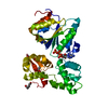
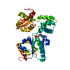
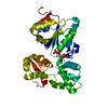
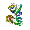
| Entry | Database: PDB / ID: 7cua | ||||||
|---|---|---|---|---|---|---|---|
| Title | The structure of YoeB dimer from Staphylococcus aureus | ||||||
 Components Components | YoeB | ||||||
 Keywords Keywords | TOXIN / toxin-antitoxin / microbial RNase / YoeB / Staphylococcus aureus | ||||||
| Function / homology | Toxin YoeB / YoeB-like toxin of bacterial type II toxin-antitoxin system / Toxin-antitoxin system, RelE/ParE toxin domain superfamily / RNA catabolic process / endonuclease activity / negative regulation of DNA-templated transcription / hydrolase activity / RNA binding / Toxin YoeB Function and homology information Function and homology information | ||||||
| Biological species |   Staphylococcus aureus (bacteria) Staphylococcus aureus (bacteria) | ||||||
| Method |  X-RAY DIFFRACTION / SYNCHROTRON / MOLECULAR REPLACEMENT / Resolution: 1.8 Å X-RAY DIFFRACTION / SYNCHROTRON / MOLECULAR REPLACEMENT / Resolution: 1.8 Å | ||||||
 Authors Authors | Yue, J. / Xue, L. | ||||||
| Funding support |  China, 1items China, 1items
| ||||||
 Citation Citation | Journal: Nucleic Acids Res. / Year: 2020 Title: Distinct oligomeric structures of the YoeB-YefM complex provide insights into the conditional cooperativity of type II toxin-antitoxin system. Authors: Xue, L. / Yue, J. / Ke, J. / Khan, M.H. / Wen, W. / Sun, B. / Zhu, Z. / Niu, L. | ||||||
| History |
|
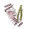
- Structure visualization
| Structure viewer | Molecule: MolmilJmol/JSmol |
|---|
- Downloads & links
-Download
| PDBx/mmCIF format | 7cua.cif.gz | 85 KB | Display | PDBx/mmCIF format |
|---|---|---|---|---|
| PDB format | pdb7cua.ent.gz | 51.4 KB | Display | PDB format |
| PDBx/mmJSON format | 7cua.json.gz | Tree view | PDBx/mmJSON format | |
| Others |  Other downloads Other downloads |
-Validation report
| Arichive directory | https://data.pdbj.org/pub/pdb/validation_reports/cu/7cuaftp://data.pdbj.org/pub/pdb/validation_reports/cu/7cua | HTTPS FTP |
|---|
-Related structure data
| Related structure data |  6l8eC  6l8fSC S: Starting model for refinement C: citing same article ( |
|---|---|
| Similar structure data |


-Links
 PDBj
PDBj- Assembly
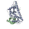
| Deposited unit | 
| ||||||||||||
|---|---|---|---|---|---|---|---|---|---|---|---|---|---|
| 1 |
| ||||||||||||
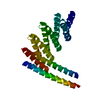
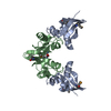
| Unit cell |
| ||||||||||||
| Components on special symmetry positions |
|
-Components
| #1: Protein | Mass: 10457.985 Da / Num. of mol.: 3 Source method: isolated from a genetically manipulated source Source: (gene. exp.) Staphylococcus aureus (strain NCTC 8325) (bacteria)Strain: NCTC 8325 / Gene: SAOUHSC_02691 / Production host: #2: Chemical |   Mass: 96.063 Da / Num. of mol.: 3 / Source method: obtained synthetically / Formula: SO4 Mass: 96.063 Da / Num. of mol.: 3 / Source method: obtained synthetically / Formula: SO4#3: Water | ChemComp-HOH / |  Mass: 18.015 Da / Num. of mol.: 198 / Source method: isolated from a natural source / Formula: H2O Mass: 18.015 Da / Num. of mol.: 198 / Source method: isolated from a natural source / Formula: H2OHas ligand of interest | N | |
|---|
-Experimental details
-Experiment
| Experiment | Method: X-RAY DIFFRACTION / Number of used crystals: 1 |
|---|
- Sample preparation
| Crystal | Density Matthews: 2.37 Å3/Da / Density % sol: 48.08 % |
|---|---|
| Crystal grow | Temperature: 289 K / Method: vapor diffusion, sitting drop Details: 25% w/v polyethylene glycol 3350, 0.1 M citric acid pH 3.5, with 1/5 volume additive of 20% 1.0 M ammonium sulfate. |
-Data collection
| Diffraction | Mean temperature: 100 K / Serial crystal experiment: N |
|---|---|
| Diffraction source | Source: SYNCHROTRON / Site: SSRF / Beamline: BL19U1 / Wavelength: 0.97852 Å |
| Detector | Type: DECTRIS PILATUS 2M / Detector: PIXEL / Date: May 24, 2020 |
| Radiation | Protocol: SINGLE WAVELENGTH / Monochromatic (M) / Laue (L): M / Scattering type: x-ray |
| Radiation wavelength | Wavelength: 0.97852 Å / Relative weight: 1 |
| Reflection | Resolution: 1.8→77.45 Å / Num. obs: 27885 / % possible obs: 99.8 % / Redundancy: 12.9 % / Biso Wilson estimate: 22.82 Å2 / CC1/2: 0.999 / Rmerge(I) obs: 0.1096 / Rpim(I) all: 0.1142 / Rrim(I) all: 0.032 / Net I/σ(I): 18.3 |
| Reflection shell | Resolution: 1.8→1.86 Å / Rmerge(I) obs: 1.199 / Num. unique obs: 2724 / CC1/2: 0.797 / Rpim(I) all: 1.247 / Rrim(I) all: 0.339 |
- Processing
| Software |
| |||||||||||||||||||||||||||||||||||||||||||||||||||||||||||||||||||||||||||||
|---|---|---|---|---|---|---|---|---|---|---|---|---|---|---|---|---|---|---|---|---|---|---|---|---|---|---|---|---|---|---|---|---|---|---|---|---|---|---|---|---|---|---|---|---|---|---|---|---|---|---|---|---|---|---|---|---|---|---|---|---|---|---|---|---|---|---|---|---|---|---|---|---|---|---|---|---|---|---|
| Refinement | Method to determine structure: MOLECULAR REPLACEMENT Starting model: 6L8F Resolution: 1.8→45.72 Å / SU ML: 0.2185 / Cross valid method: FREE R-VALUE / σ(F): 1.34 / Phase error: 23.1867 Stereochemistry target values: GeoStd + Monomer Library + CDL v1.2
| |||||||||||||||||||||||||||||||||||||||||||||||||||||||||||||||||||||||||||||
| Solvent computation | Shrinkage radii: 0.9 Å / VDW probe radii: 1.11 Å / Solvent model: FLAT BULK SOLVENT MODEL | |||||||||||||||||||||||||||||||||||||||||||||||||||||||||||||||||||||||||||||
| Displacement parameters | Biso mean: 25.7 Å2 | |||||||||||||||||||||||||||||||||||||||||||||||||||||||||||||||||||||||||||||
| Refinement step | Cycle: LAST / Resolution: 1.8→45.72 Å
| |||||||||||||||||||||||||||||||||||||||||||||||||||||||||||||||||||||||||||||
| Refine LS restraints |
| |||||||||||||||||||||||||||||||||||||||||||||||||||||||||||||||||||||||||||||
| LS refinement shell |
|
