| 登録情報 | データベース: PDB / ID: 4xlg
|
|---|
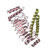
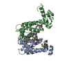
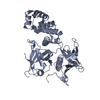
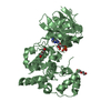
| タイトル | C. glabrata Slx1 in complex with Slx4CCD. |
|---|
 要素 要素 | - Structure-specific endonuclease subunit SLX1
- Structure-specific endonuclease subunit SLX4
|
|---|
 キーワード キーワード | HYDROLASE / nuclease / DNA repair / GIY-YIG / homogolous recombination |
|---|
| 機能・相同性 |  機能・相同性情報 機能・相同性情報
Slx1-Slx4 complex / crossover junction DNA endonuclease activity / 5'-flap endonuclease activity / DNA-templated DNA replication / double-strand break repair via homologous recombination / DNA recombination / 加水分解酵素; エステル加水分解酵素 / DNA replication / DNA repair / zinc ion binding類似検索 - 分子機能 Structure-specific endonuclease subunit Slx4, ascomycetes / : / Structure-specific endonuclease subunit SLX1, C-terminal / Structure-specific endonuclease subunit Slx1 / : / Structure-specific endonuclease subunit Slx4 / Slx4 endonuclease / GIY-YIG type nucleases (URI domain) / GIY-YIG endonuclease superfamily / GIY-YIG catalytic domain ...Structure-specific endonuclease subunit Slx4, ascomycetes / : / Structure-specific endonuclease subunit SLX1, C-terminal / Structure-specific endonuclease subunit Slx1 / : / Structure-specific endonuclease subunit Slx4 / Slx4 endonuclease / GIY-YIG type nucleases (URI domain) / GIY-YIG endonuclease superfamily / GIY-YIG catalytic domain / GIY-YIG endonuclease / GIY-YIG domain profile. / Zinc finger, RING/FYVE/PHD-type類似検索 - ドメイン・相同性 Structure-specific endonuclease subunit SLX4 / Structure-specific endonuclease subunit SLX1類似検索 - 構成要素 |
|---|
| 生物種 |  Candida glabrata (菌類) Candida glabrata (菌類) |
|---|
| 手法 |  X線回折 / シンクロトロン / 分子置換 / 解像度: 1.78 Å X線回折 / シンクロトロン / 分子置換 / 解像度: 1.78 Å |
|---|
 データ登録者 データ登録者 | Gaur, V. / Wyatt, H.D.M. / Komorowska, W. / Szczepanowski, R.H. / de Sanctis, D. / Gorecka, K.M. / West, S.C. / Nowotny, M. |
|---|
| 資金援助 |  英国, 1件 英国, 1件 | 組織 | 認可番号 | 国 |
|---|
| Wellcome Trust | 98022 | 英国 |
|
|---|
 引用 引用 | ジャーナル: Cell Rep / 年: 2015
タイトル: Structural and Mechanistic Analysis of the Slx1-Slx4 Endonuclease.
著者: Gaur, V. / Wyatt, H.D. / Komorowska, W. / Szczepanowski, R.H. / de Sanctis, D. / Gorecka, K.M. / West, S.C. / Nowotny, M. |
|---|
| 履歴 | | 登録 | 2015年1月13日 | 登録サイト: RCSB / 処理サイト: PDBE |
|---|
| 改定 1.0 | 2015年3月25日 | Provider: repository / タイプ: Initial release |
|---|
| 改定 1.1 | 2015年5月20日 | Group: Database references |
|---|
| 改定 1.2 | 2024年1月10日 | Group: Data collection / Database references / Refinement description
カテゴリ: chem_comp_atom / chem_comp_bond ...chem_comp_atom / chem_comp_bond / database_2 / pdbx_initial_refinement_model
Item: _database_2.pdbx_DOI / _database_2.pdbx_database_accession |
|---|
|
|---|
 ムービー
ムービー コントローラー


 データを開く
 基本情報
基本情報 構造の表示
構造の表示 ダウンロードとリンク
 その他のダウンロード


 PDBj
PDBj 集合体
集合体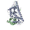


 分子量: 65.409 Da / 分子数: 2 / 由来タイプ: 合成 / 式: Zn
 分子量: 35.453 Da / 分子数: 2 / 由来タイプ: 合成 / 式: Cl
 分子量: 18.015 Da / 分子数: 302 / 由来タイプ: 天然 / 式: H2O
 試料調製
試料調製 / ビームライン: ID29 / 波長: 0.97626 Å
 解析
解析