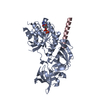
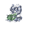
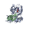
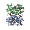
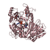
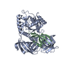
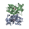
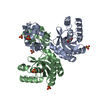
登録情報 データベース : PDB / ID : 7cslタイトル Crystal structure of the archaeal EF1A-EF1B complex Elongation factor 1-alpha Elongation factor 1-beta キーワード / / 機能・相同性 分子機能 ドメイン・相同性 構成要素
/ / / / / / / / / / / / / / / / / / / / / / / / / / / / / / / / / / / / / / / / / / 生物種 Pyrococcus horikoshii OT-3 (古細菌)手法 / / / 解像度 : 2 Å データ登録者 Suzuki, T. / Ito, K. / Miyoshi, T. / Murakami, R. / Uchiumi, T. 資金援助 組織 認可番号 国 Japan Society for the Promotion of Science (JSPS) 19H03155
ジャーナル : J.Mol.Biol. / 年 : 2021タイトル : Structural insights into the Switching Off of the Interaction between the Archaeal Ribosomal Stalk and aEF1A by Nucleotide Exchange Factor aEF1B.著者 : Suzuki, T. / Ito, K. / Miyoshi, T. / Murakami, R. / Uchiumi, T. 履歴 登録 2020年8月15日 登録サイト / 処理サイト 改定 1.0 2021年6月23日 Provider / タイプ 改定 1.1 2021年7月28日 Group / カテゴリ / Item 改定 1.2 2023年11月29日 Group / Database references / Refinement descriptionカテゴリ chem_comp_atom / chem_comp_bond ... chem_comp_atom / chem_comp_bond / database_2 / pdbx_initial_refinement_model Item / _database_2.pdbx_database_accession
すべて表示 表示を減らす
 ムービー
ムービー コントローラー


 データを開く
 基本情報
基本情報 要素
要素 キーワード
キーワード 機能・相同性情報
 Pyrococcus horikoshii OT-3 (古細菌)
 X線回折 /
  データ登録者
 日本, 1件
  引用
引用 構造の表示
構造の表示 ダウンロードとリンク
 その他のダウンロード
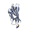


 PDBj
PDBj


 集合体
集合体


 分子量: 18.015 Da / 分子数: 251 / 由来タイプ: 天然 / 式: H2O
 試料調製
試料調製 解析
解析