 Movie
Movie Controller

[English] 日本語
 Yorodumi
- PDB-7cmj: Crystal structure of L.donovani Hypoxanthine-guanine phosphoribos... -
+ Open data

- Basic information
| Entry | Database: PDB / ID: 7cmj | ||||||
|---|---|---|---|---|---|---|---|
| Title | Crystal structure of L.donovani Hypoxanthine-guanine phosphoribosyl transferase (HGPRT) | ||||||
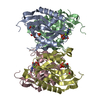
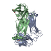
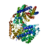
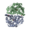
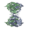
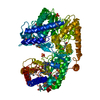
 Components Components | Hypoxanthine phosphoribosyltransferase | ||||||
 Keywords Keywords | TRANSFERASE / hypoxanthine / guanine / phosphoribosyl / complex / tetramer. | ||||||
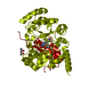
| Function / homology |  Function and homology information Function and homology informationhypoxanthine phosphoribosyltransferase / guanine phosphoribosyltransferase activity / hypoxanthine phosphoribosyltransferase activity / IMP salvage / purine ribonucleoside salvage / nucleotide binding / metal ion binding / cytoplasm Similarity search - Function | ||||||
| Biological species |  Leishmania donovani (eukaryote) Leishmania donovani (eukaryote) | ||||||
| Method |  X-RAY DIFFRACTION / SYNCHROTRON / MOLECULAR REPLACEMENT / Resolution: 2.76 Å X-RAY DIFFRACTION / SYNCHROTRON / MOLECULAR REPLACEMENT / Resolution: 2.76 Å | ||||||
 Authors Authors | Parihar, P.S. / Pratap, J.V. | ||||||
 Citation Citation | Journal: Biochem.Biophys.Res.Commun. / Year: 2020 Title: The L.donovani Hypoxanthine-guanine phosphoribosyl transferase (HGPRT) oligomer is distinct from the human homolog. Authors: Parihar, P.S. / Pratap, J.V. | ||||||
| History |
|
- Structure visualization
| Structure viewer | Molecule: MolmilJmol/JSmol |
|---|
- Downloads & links
-Download
| PDBx/mmCIF format | 7cmj.cif.gz | 161.2 KB | Display | PDBx/mmCIF format |
|---|---|---|---|---|
| PDB format | pdb7cmj.ent.gz | 125.8 KB | Display | PDB format |
| PDBx/mmJSON format | 7cmj.json.gz | Tree view | PDBx/mmJSON format | |
| Others |  Other downloads Other downloads |
-Validation report
| Arichive directory | https://data.pdbj.org/pub/pdb/validation_reports/cm/7cmjftp://data.pdbj.org/pub/pdb/validation_reports/cm/7cmj | HTTPS FTP |
|---|
-Related structure data
| Related structure data |  1pzmS S: Starting model for refinement |
|---|---|
| Similar structure data |


-Links
 PDBj
PDBj
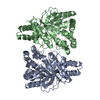
- Assembly
| Deposited unit | 
| ||||||||
|---|---|---|---|---|---|---|---|---|---|
| 1 | 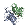
| ||||||||
| Unit cell |
|
-Components
-Protein , 1 types, 2 molecules AB
| #1: Protein | Mass: 23642.365 Da / Num. of mol.: 2 Source method: isolated from a genetically manipulated source Details: unlined residue have missing electron density, side chain of some residue also missing are with removed side chain. Source: (gene. exp.) Leishmania donovani (strain BPK282A1) (eukaryote)Strain: BPK282A1 / Gene: LDBPK_210980 / Production host:  References: UniProt: E9BF84, hypoxanthine phosphoribosyltransferase |
|---|
-Non-polymers , 7 types, 120 molecules 


| #2: Chemical |  Mass: 24.305 Da / Num. of mol.: 2 / Source method: obtained synthetically / Formula: Mg / Feature type: SUBJECT OF INVESTIGATION Mass: 24.305 Da / Num. of mol.: 2 / Source method: obtained synthetically / Formula: Mg / Feature type: SUBJECT OF INVESTIGATION#3: Chemical |  Mass: 137.327 Da / Num. of mol.: 2 / Source method: obtained synthetically / Formula: Ba / Feature type: SUBJECT OF INVESTIGATION Mass: 137.327 Da / Num. of mol.: 2 / Source method: obtained synthetically / Formula: Ba / Feature type: SUBJECT OF INVESTIGATION#4: Chemical |  Mass: 94.971 Da / Num. of mol.: 2 / Source method: obtained synthetically / Formula: PO4 / Feature type: SUBJECT OF INVESTIGATION Mass: 94.971 Da / Num. of mol.: 2 / Source method: obtained synthetically / Formula: PO4 / Feature type: SUBJECT OF INVESTIGATION#5: Chemical |  Mass: 106.120 Da / Num. of mol.: 2 / Source method: obtained synthetically / Formula: C4H10O3 / Feature type: SUBJECT OF INVESTIGATION Mass: 106.120 Da / Num. of mol.: 2 / Source method: obtained synthetically / Formula: C4H10O3 / Feature type: SUBJECT OF INVESTIGATION#6: Chemical | ChemComp-PG4 / |  Mass: 194.226 Da / Num. of mol.: 1 / Source method: obtained synthetically / Formula: C8H18O5 / Feature type: SUBJECT OF INVESTIGATION / Comment: precipitant*YM Mass: 194.226 Da / Num. of mol.: 1 / Source method: obtained synthetically / Formula: C8H18O5 / Feature type: SUBJECT OF INVESTIGATION / Comment: precipitant*YM#7: Chemical |  Mass: 35.453 Da / Num. of mol.: 2 / Source method: obtained synthetically / Formula: Cl / Feature type: SUBJECT OF INVESTIGATION Mass: 35.453 Da / Num. of mol.: 2 / Source method: obtained synthetically / Formula: Cl / Feature type: SUBJECT OF INVESTIGATION#8: Water | ChemComp-HOH / | Mass: 18.015 Da / Num. of mol.: 109 / Source method: isolated from a natural source / Formula: H2O |
|---|
-Details
| Has ligand of interest | Y |
|---|
-Experimental details
-Experiment
| Experiment | Method: X-RAY DIFFRACTION / Number of used crystals: 1 |
|---|
- Sample preparation
| Crystal | Density Matthews: 3.64 Å3/Da / Density % sol: 70.39 % |
|---|---|
| Crystal grow | Temperature: 288 K / Method: vapor diffusion, hanging drop / pH: 6.5 Details: 0.1M Sodium cacodylate, 0.2 M Ammonium sulfate, 20% PEG 4000 PH range: 6-7 |
-Data collection
| Diffraction | Mean temperature: 100 K / Serial crystal experiment: N |
|---|---|
| Diffraction source | Source: SYNCHROTRON / Site: ELETTRA  / Beamline: 11.2C / Wavelength: 0.9784 Å / Beamline: 11.2C / Wavelength: 0.9784 Å |
| Detector | Type: DECTRIS PILATUS 6M / Detector: PIXEL / Date: Feb 13, 2019 / Details: Monochromator |
| Radiation | Protocol: SINGLE WAVELENGTH / Monochromatic (M) / Laue (L): M / Scattering type: x-ray |
| Radiation wavelength | Wavelength: 0.9784 Å / Relative weight: 1 |
| Reflection | Resolution: 2.76→44.373 Å / Num. obs: 17049 / % possible obs: 93.7 % / Redundancy: 1.9 % / Biso Wilson estimate: 52.65 Å2 / Rmerge(I) obs: 0.04166 / Net I/σ(I): 10.15 |
| Reflection shell | Resolution: 2.76→2.86 Å / Redundancy: 1.8 % / Rmerge(I) obs: 0.2919 / Num. unique obs: 1731 / % possible all: 98.1 |
- Processing
| Software |
| |||||||||||||||||||||||||||||||||||||||||||||||||
|---|---|---|---|---|---|---|---|---|---|---|---|---|---|---|---|---|---|---|---|---|---|---|---|---|---|---|---|---|---|---|---|---|---|---|---|---|---|---|---|---|---|---|---|---|---|---|---|---|---|---|
| Refinement | Method to determine structure: MOLECULAR REPLACEMENT Starting model: 1PZM Resolution: 2.76→44.37 Å / SU ML: 0.34 / Cross valid method: FREE R-VALUE / σ(F): 1.34 / Phase error: 22.91 / Stereochemistry target values: ML
| |||||||||||||||||||||||||||||||||||||||||||||||||
| Solvent computation | Shrinkage radii: 0.9 Å / VDW probe radii: 1.11 Å / Solvent model: FLAT BULK SOLVENT MODEL | |||||||||||||||||||||||||||||||||||||||||||||||||
| Refinement step | Cycle: LAST / Resolution: 2.76→44.37 Å
| |||||||||||||||||||||||||||||||||||||||||||||||||
| Refine LS restraints |
| |||||||||||||||||||||||||||||||||||||||||||||||||
| LS refinement shell |
| |||||||||||||||||||||||||||||||||||||||||||||||||
| Refinement TLS params. | Method: refined / Origin x: -31.4031 Å / Origin y: 31.0988 Å / Origin z: -11.5201 Å
| |||||||||||||||||||||||||||||||||||||||||||||||||
| Refinement TLS group | Selection details: ALL |
