 Movie
Movie Controller

[English] 日本語
 Yorodumi
- PDB-5kny: Crystal structure of Mycobacterium tuberculosis hypoxanthine guan... -
+ Open data

- Basic information
| Entry | Database: PDB / ID: 5kny | ||||||
|---|---|---|---|---|---|---|---|
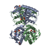
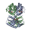
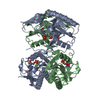
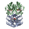
| Title | Crystal structure of Mycobacterium tuberculosis hypoxanthine guanine phosphoribosyltransferase in complex with (3-((3R,4R)-3-(Guanin-9-yl)-4-((S)-2-hydroxy-2-phosphonoethoxy)pyrrolidin-1-yl)-3-oxopropyl)phosphonic acid | ||||||
 Components Components | Hypoxanthine-guanine phosphoribosyltransferase | ||||||
 Keywords Keywords | TRANSFERASE/INHIBITOR / Phosphoribosyltransferase / Inhibitor / Complex / pyrrolidne nucleoside phosphonate / TRANSFERASE-INHIBITOR complex | ||||||
| Function / homology |  Function and homology information Function and homology informationIMP biosynthetic process / purine-containing compound salvage / hypoxanthine phosphoribosyltransferase / guanine phosphoribosyltransferase activity / guanine salvage / hypoxanthine metabolic process / hypoxanthine phosphoribosyltransferase activity / GMP salvage / IMP salvage / GMP biosynthetic process ...IMP biosynthetic process / purine-containing compound salvage / hypoxanthine phosphoribosyltransferase / guanine phosphoribosyltransferase activity / guanine salvage / hypoxanthine metabolic process / hypoxanthine phosphoribosyltransferase activity / GMP salvage / IMP salvage / GMP biosynthetic process / purine ribonucleoside salvage / nucleotide binding / magnesium ion binding / cytosol Similarity search - Function | ||||||
| Biological species |   Mycobacterium tuberculosis (bacteria) Mycobacterium tuberculosis (bacteria) | ||||||
| Method |  X-RAY DIFFRACTION / SYNCHROTRON / Resolution: 2.91 Å X-RAY DIFFRACTION / SYNCHROTRON / Resolution: 2.91 Å | ||||||
 Authors Authors | Eng, W.S. / Rejman, D. / Keough, D.T. / Guddat, L.W. | ||||||
 Citation Citation | Journal: To Be Published Title: Crystal structure of Mycobacterium tuberculosis hypoxanthine guanine phosphoribosyltransferase in complex with pyrrolidine nucleoside phosphonate Authors: Eng, W.S. / Rejman, D. / Keough, D.T. / Guddat, L.W. | ||||||
| History |
|
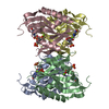
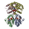
- Structure visualization
| Structure viewer | Molecule: MolmilJmol/JSmol |
|---|
- Downloads & links
-Download
| PDBx/mmCIF format | 5kny.cif.gz | 295.5 KB | Display | PDBx/mmCIF format |
|---|---|---|---|---|
| PDB format | pdb5kny.ent.gz | 241.1 KB | Display | PDB format |
| PDBx/mmJSON format | 5kny.json.gz | Tree view | PDBx/mmJSON format | |
| Others |  Other downloads Other downloads |
-Validation report
| Arichive directory | https://data.pdbj.org/pub/pdb/validation_reports/kn/5knyftp://data.pdbj.org/pub/pdb/validation_reports/kn/5kny | HTTPS FTP |
|---|
-Related structure data


-Links
 PDBj
PDBj
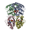
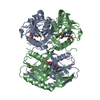
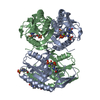
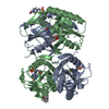
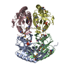
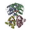
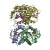
- Assembly
| Deposited unit | 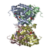
| |||||||||||||||||||||||||||||||||||||||||||||
|---|---|---|---|---|---|---|---|---|---|---|---|---|---|---|---|---|---|---|---|---|---|---|---|---|---|---|---|---|---|---|---|---|---|---|---|---|---|---|---|---|---|---|---|---|---|---|
| 1 |
| |||||||||||||||||||||||||||||||||||||||||||||
| 2 | 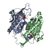
| |||||||||||||||||||||||||||||||||||||||||||||
| 3 | 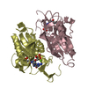
| |||||||||||||||||||||||||||||||||||||||||||||
| Unit cell |
| |||||||||||||||||||||||||||||||||||||||||||||
| Noncrystallographic symmetry (NCS) | NCS domain:
NCS domain segments:
|
-Components
| #1: Protein | Mass: 22969.801 Da / Num. of mol.: 4 Source method: isolated from a genetically manipulated source Source: (gene. exp.) Mycobacterium tuberculosis (strain ATCC 25618 / H37Rv) (bacteria)Strain: ATCC 25618 / H37Rv / Gene: hpt, hprT, Rv3624c, MTCY15C10.28 / Production host: References: UniProt: P9WHQ9, hypoxanthine phosphoribosyltransferase #2: Chemical | ChemComp-YPG / [ 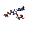  Mass: 496.306 Da / Num. of mol.: 4 / Source method: obtained synthetically / Formula: C14H22N6O10P2 Mass: 496.306 Da / Num. of mol.: 4 / Source method: obtained synthetically / Formula: C14H22N6O10P2#3: Chemical | ChemComp-MG /   Mass: 24.305 Da / Num. of mol.: 4 / Source method: obtained synthetically / Formula: Mg Mass: 24.305 Da / Num. of mol.: 4 / Source method: obtained synthetically / Formula: Mg#4: Water | ChemComp-HOH / |  Mass: 18.015 Da / Num. of mol.: 29 / Source method: isolated from a natural source / Formula: H2O Mass: 18.015 Da / Num. of mol.: 29 / Source method: isolated from a natural source / Formula: H2O |
|---|
-Experimental details
-Experiment
| Experiment | Method: X-RAY DIFFRACTION / Number of used crystals: 1 |
|---|
- Sample preparation
| Crystal | Density Matthews: 1.96 Å3/Da / Density % sol: 37.25 % |
|---|---|
| Crystal grow | Temperature: 291 K / Method: vapor diffusion, hanging drop Details: 0.2 M Li2SO4, 0.1 M Sodium acetate pH 4.5 and 30% PEG8000 |
-Data collection
| Diffraction | Mean temperature: 100 K |
|---|---|
| Diffraction source | Source: SYNCHROTRON / Site: Australian Synchrotron  / Beamline: MX1 / Wavelength: 0.9537 Å / Beamline: MX1 / Wavelength: 0.9537 Å |
| Detector | Type: ADSC QUANTUM 270 / Detector: CCD / Date: Jun 27, 2014 |
| Radiation | Protocol: SINGLE WAVELENGTH / Monochromatic (M) / Laue (L): M / Scattering type: x-ray |
| Radiation wavelength | Wavelength: 0.9537 Å / Relative weight: 1 |
| Reflection | Resolution: 2.91→44.77 Å / Num. obs: 15646 / % possible obs: 99.8 % / Redundancy: 3.8 % / Net I/σ(I): 7.8 |
| Reflection shell | Resolution: 2.91→3.09 Å / Redundancy: 3.8 % / Rmerge(I) obs: 0.587 / Mean I/σ(I) obs: 1.8 / % possible all: 99.4 |
- Processing
| Software |
| |||||||||||||||||||||||||||||||||||||||||||||||||||||||||||||||||||||||||||||||||||||||||||||||||||||||||||||||||||||||||||||
|---|---|---|---|---|---|---|---|---|---|---|---|---|---|---|---|---|---|---|---|---|---|---|---|---|---|---|---|---|---|---|---|---|---|---|---|---|---|---|---|---|---|---|---|---|---|---|---|---|---|---|---|---|---|---|---|---|---|---|---|---|---|---|---|---|---|---|---|---|---|---|---|---|---|---|---|---|---|---|---|---|---|---|---|---|---|---|---|---|---|---|---|---|---|---|---|---|---|---|---|---|---|---|---|---|---|---|---|---|---|---|---|---|---|---|---|---|---|---|---|---|---|---|---|---|---|---|
| Refinement | Resolution: 2.91→44.77 Å / SU ML: 0.39 / Cross valid method: FREE R-VALUE / σ(F): 1.37 / Phase error: 26.22
| |||||||||||||||||||||||||||||||||||||||||||||||||||||||||||||||||||||||||||||||||||||||||||||||||||||||||||||||||||||||||||||
| Solvent computation | Shrinkage radii: 0.9 Å / VDW probe radii: 1.11 Å | |||||||||||||||||||||||||||||||||||||||||||||||||||||||||||||||||||||||||||||||||||||||||||||||||||||||||||||||||||||||||||||
| Displacement parameters | Biso max: 136.95 Å2 / Biso mean: 53.2634 Å2 / Biso min: 12.65 Å2 | |||||||||||||||||||||||||||||||||||||||||||||||||||||||||||||||||||||||||||||||||||||||||||||||||||||||||||||||||||||||||||||
| Refinement step | Cycle: final / Resolution: 2.91→44.77 Å
| |||||||||||||||||||||||||||||||||||||||||||||||||||||||||||||||||||||||||||||||||||||||||||||||||||||||||||||||||||||||||||||
| Refine LS restraints |
| |||||||||||||||||||||||||||||||||||||||||||||||||||||||||||||||||||||||||||||||||||||||||||||||||||||||||||||||||||||||||||||
| Refine LS restraints NCS |
| |||||||||||||||||||||||||||||||||||||||||||||||||||||||||||||||||||||||||||||||||||||||||||||||||||||||||||||||||||||||||||||
| LS refinement shell | Refine-ID: X-RAY DIFFRACTION / Total num. of bins used: 11
| |||||||||||||||||||||||||||||||||||||||||||||||||||||||||||||||||||||||||||||||||||||||||||||||||||||||||||||||||||||||||||||
| Refinement TLS params. | Method: refined / Refine-ID: X-RAY DIFFRACTION
| |||||||||||||||||||||||||||||||||||||||||||||||||||||||||||||||||||||||||||||||||||||||||||||||||||||||||||||||||||||||||||||
| Refinement TLS group |
|
