 Movie
Movie Controller

[English] 日本語
 Yorodumi
- PDB-7cay: Crystal Structure of Lon N-terminal domain protein from Xanthomon... -
+ Open data

- Basic information
| Entry | Database: PDB / ID: 7cay | ||||||
|---|---|---|---|---|---|---|---|
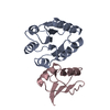
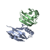
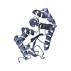
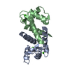
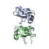
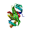
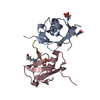
| Title | Crystal Structure of Lon N-terminal domain protein from Xanthomonas campestris | ||||||
 Components Components | ATP-dependent protease | ||||||
 Keywords Keywords | PROTEIN BINDING / N-terminal domain / Lon protease | ||||||
| Function / homology |  Function and homology information Function and homology information | ||||||
| Biological species |  Xanthomonas campestris pv. campestris (bacteria) Xanthomonas campestris pv. campestris (bacteria) | ||||||
| Method |  X-RAY DIFFRACTION / SYNCHROTRON / MOLECULAR REPLACEMENT / Resolution: 2.8 Å X-RAY DIFFRACTION / SYNCHROTRON / MOLECULAR REPLACEMENT / Resolution: 2.8 Å | ||||||
 Authors Authors | Singh, R. / Sharma, B. / Deshmukh, S. / Kumar, A. / Makde, R.D. | ||||||
 Citation Citation | Journal: Acta Crystallogr.,Sect.F / Year: 2020 Title: Crystal structure of XCC3289 from Xanthomonas campestris: homology with the N-terminal substrate-binding domain of Lon peptidase. Authors: Singh, R. / Deshmukh, S. / Kumar, A. / Goyal, V.D. / Makde, R.D. | ||||||
| History |
|
- Structure visualization
| Structure viewer | Molecule: MolmilJmol/JSmol |
|---|
- Downloads & links
-Download
| PDBx/mmCIF format | 7cay.cif.gz | 78.1 KB | Display | PDBx/mmCIF format |
|---|---|---|---|---|
| PDB format | pdb7cay.ent.gz | 57.2 KB | Display | PDB format |
| PDBx/mmJSON format | 7cay.json.gz | Tree view | PDBx/mmJSON format | |
| Others |  Other downloads Other downloads |
-Validation report
| Arichive directory | https://data.pdbj.org/pub/pdb/validation_reports/ca/7cayftp://data.pdbj.org/pub/pdb/validation_reports/ca/7cay | HTTPS FTP |
|---|
-Related structure data
| Related structure data | 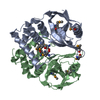 1zboS S: Starting model for refinement |
|---|---|
| Similar structure data |


-Links
 PDBj
PDBj
- Assembly
| Deposited unit | 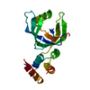
| ||||||||
|---|---|---|---|---|---|---|---|---|---|
| 1 |
| ||||||||
| Unit cell |
|
-Components
| #1: Protein | Mass: 21318.295 Da / Num. of mol.: 1 Source method: isolated from a genetically manipulated source Source: (gene. exp.) Xanthomonas campestris pv. campestris (bacteria)Gene: D0A42_17175 / Plasmid: pST50Tr / Production host: |
|---|
-Experimental details
-Experiment
| Experiment | Method: X-RAY DIFFRACTION / Number of used crystals: 1 |
|---|
- Sample preparation
| Crystal | Density Matthews: 2.54 Å3/Da / Density % sol: 51.64 % |
|---|---|
| Crystal grow | Temperature: 277 K / Method: microbatch / pH: 8.5 Details: Isopropanol 35%, 200mM ammonium acetate, 100 mM Tris-Cl, pH 8.5 |
-Data collection
| Diffraction | Mean temperature: 100 K / Serial crystal experiment: N | ||||||||||||||||||||||||||||||
|---|---|---|---|---|---|---|---|---|---|---|---|---|---|---|---|---|---|---|---|---|---|---|---|---|---|---|---|---|---|---|---|
| Diffraction source | Source: SYNCHROTRON / Site: RRCAT INDUS-2  / Beamline: PX-BL21 / Wavelength: 0.97949 Å / Beamline: PX-BL21 / Wavelength: 0.97949 Å | ||||||||||||||||||||||||||||||
| Detector | Type: RAYONIX MX-225 / Detector: CCD / Date: Mar 7, 2016 / Details: mirror | ||||||||||||||||||||||||||||||
| Radiation | Monochromator: Si111 / Protocol: SINGLE WAVELENGTH / Monochromatic (M) / Laue (L): M / Scattering type: x-ray | ||||||||||||||||||||||||||||||
| Radiation wavelength | Wavelength: 0.97949 Å / Relative weight: 1 | ||||||||||||||||||||||||||||||
| Reflection | Resolution: 2.8→44.17 Å / Num. obs: 5595 / % possible obs: 99 % / Redundancy: 8.2 % / Biso Wilson estimate: 74.2 Å2 / CC1/2: 0.999 / Rmerge(I) obs: 0.076 / Rpim(I) all: 0.028 / Rrim(I) all: 0.081 / Net I/σ(I): 19.9 / Num. measured all: 46030 / Scaling rejects: 1 | ||||||||||||||||||||||||||||||
| Reflection shell | Diffraction-ID: 1
|
- Processing
| Software |
| ||||||||||||||||||||||||||||||||||||||||
|---|---|---|---|---|---|---|---|---|---|---|---|---|---|---|---|---|---|---|---|---|---|---|---|---|---|---|---|---|---|---|---|---|---|---|---|---|---|---|---|---|---|
| Refinement | Method to determine structure: MOLECULAR REPLACEMENT Starting model: 1zbo Resolution: 2.8→33.327 Å / SU ML: 0.26 / Cross valid method: THROUGHOUT / σ(F): 1.35 / Phase error: 33.08 / Stereochemistry target values: ML
| ||||||||||||||||||||||||||||||||||||||||
| Solvent computation | Shrinkage radii: 0.9 Å / VDW probe radii: 1.11 Å / Solvent model: FLAT BULK SOLVENT MODEL | ||||||||||||||||||||||||||||||||||||||||
| Displacement parameters | Biso max: 104.63 Å2 / Biso mean: 71.1476 Å2 / Biso min: 48.99 Å2 | ||||||||||||||||||||||||||||||||||||||||
| Refinement step | Cycle: final / Resolution: 2.8→33.327 Å
| ||||||||||||||||||||||||||||||||||||||||
| Refine LS restraints |
| ||||||||||||||||||||||||||||||||||||||||
| LS refinement shell | Refine-ID: X-RAY DIFFRACTION / Rfactor Rfree error: 0 / Num. reflection Rfree: 160
| ||||||||||||||||||||||||||||||||||||||||
| Refinement TLS params. | Method: refined / Origin x: -30.9683 Å / Origin y: 11.2254 Å / Origin z: -6.3041 Å
| ||||||||||||||||||||||||||||||||||||||||
| Refinement TLS group | Selection details: all |
