 Movie
Movie Controller

[English] 日本語
 Yorodumi
- PDB-7c8j: Structural basis for cross-species recognition of COVID-19 virus ... -
+ Open data

- Basic information
| Entry | Database: PDB / ID: 7c8j | ||||||
|---|---|---|---|---|---|---|---|
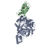
| Title | Structural basis for cross-species recognition of COVID-19 virus spike receptor binding domain to bat ACE2 | ||||||
 Components Components |
| ||||||
 Keywords Keywords | VIRAL PROTEIN/PROTEIN BINDING / COVID-19 / receptor binding domain (RBD) / Rhinolophus macrotis / bats / ACE2 / VIRAL PROTEIN-PROTEIN BINDING complex | ||||||
| Function / homology |  Function and homology information Function and homology informationHydrolases; Acting on peptide bonds (peptidases) / peptidyl-dipeptidase activity / carboxypeptidase activity / metallopeptidase activity / symbiont-mediated disruption of host tissue / Maturation of spike protein / Translation of Structural Proteins / Virion Assembly and Release / host cell surface / host extracellular region ...Hydrolases; Acting on peptide bonds (peptidases) / peptidyl-dipeptidase activity / carboxypeptidase activity / metallopeptidase activity / symbiont-mediated disruption of host tissue / Maturation of spike protein / Translation of Structural Proteins / Virion Assembly and Release / host cell surface / host extracellular region / symbiont-mediated-mediated suppression of host tetherin activity / Induction of Cell-Cell Fusion / structural constituent of virion / positive regulation of viral entry into host cell / membrane fusion / host cell endoplasmic reticulum-Golgi intermediate compartment membrane / Attachment and Entry / entry receptor-mediated virion attachment to host cell / receptor-mediated virion attachment to host cell / apical plasma membrane / host cell surface receptor binding / symbiont-mediated suppression of host innate immune response / cilium / endocytosis involved in viral entry into host cell / receptor ligand activity / fusion of virus membrane with host plasma membrane / fusion of virus membrane with host endosome membrane / viral envelope / symbiont entry into host cell / virion attachment to host cell / host cell plasma membrane / SARS-CoV-2 activates/modulates innate and adaptive immune responses / virion membrane / proteolysis / : / membrane / metal ion binding / identical protein binding / plasma membrane / cytoplasm Similarity search - Function | ||||||
| Biological species |  Rhinolophus macrotis (Big-eared Horseshoe Bat) Rhinolophus macrotis (Big-eared Horseshoe Bat)  Severe acute respiratory syndrome coronavirus 2 Severe acute respiratory syndrome coronavirus 2 | ||||||
| Method |  X-RAY DIFFRACTION / SYNCHROTRON / MOLECULAR REPLACEMENT / Resolution: 3.18 Å X-RAY DIFFRACTION / SYNCHROTRON / MOLECULAR REPLACEMENT / Resolution: 3.18 Å | ||||||
 Authors Authors | Liu, K.F. / Wang, J. / Tan, S.G. / Niu, S. / Wu, L.L. / Zhang, Y.F. / Pan, X.Q. / Meng, Y.M. / Chen, Q. / Wang, Q.H. ...Liu, K.F. / Wang, J. / Tan, S.G. / Niu, S. / Wu, L.L. / Zhang, Y.F. / Pan, X.Q. / Meng, Y.M. / Chen, Q. / Wang, Q.H. / Wang, H.W. / Qi, J.X. / Gao, G.F. | ||||||
 Citation Citation | Journal: Proc Natl Acad Sci U S A / Year: 2021 Title: Cross-species recognition of SARS-CoV-2 to bat ACE2. Authors: Kefang Liu / Shuguang Tan / Sheng Niu / Jia Wang / Lili Wu / Huan Sun / Yanfang Zhang / Xiaoqian Pan / Xiao Qu / Pei Du / Yumin Meng / Yunfei Jia / Qian Chen / Chuxia Deng / Jinghua Yan / ...Authors: Kefang Liu / Shuguang Tan / Sheng Niu / Jia Wang / Lili Wu / Huan Sun / Yanfang Zhang / Xiaoqian Pan / Xiao Qu / Pei Du / Yumin Meng / Yunfei Jia / Qian Chen / Chuxia Deng / Jinghua Yan / Hong-Wei Wang / Qihui Wang / Jianxun Qi / George Fu Gao /  Abstract: The coronavirus disease 2019 (COVID-19) pandemic caused by severe acute respiratory syndrome coronavirus 2 (SARS-CoV-2) has emerged as a major threat to global health. Although varied SARS-CoV-2- ...The coronavirus disease 2019 (COVID-19) pandemic caused by severe acute respiratory syndrome coronavirus 2 (SARS-CoV-2) has emerged as a major threat to global health. Although varied SARS-CoV-2-related coronaviruses have been isolated from bats and SARS-CoV-2 may infect bat, the structural basis for SARS-CoV-2 to utilize the human receptor counterpart bat angiotensin-converting enzyme 2 (bACE2) for virus infection remains less understood. Here, we report that the SARS-CoV-2 spike protein receptor binding domain (RBD) could bind to bACE2 from (bACE2-Rm) with substantially lower affinity compared with that to the human ACE2 (hACE2), and its infectivity to host cells expressing bACE2-Rm was confirmed with pseudotyped SARS-CoV-2 virus and SARS-CoV-2 wild virus. The structure of the SARS-CoV-2 RBD with the bACE2-Rm complex was determined, revealing a binding mode similar to that of hACE2. The analysis of binding details between SARS-CoV-2 RBD and bACE2-Rm revealed that the interacting network involving Y41 and E42 of bACE2-Rm showed substantial differences with that to hACE2. Bats have extensive species diversity and the residues for RBD binding in bACE2 receptor varied substantially among different bat species. Notably, the Y41H mutant, which exists in many bats, attenuates the binding capacity of bACE2-Rm, indicating the central roles of Y41 in the interaction network. These findings would benefit our understanding of the potential infection of SARS-CoV-2 in varied species of bats. | ||||||
| History |
|
- Structure visualization
| Structure viewer | Molecule: MolmilJmol/JSmol |
|---|
- Downloads & links
-Download
| PDBx/mmCIF format | 7c8j.cif.gz | 418.7 KB | Display | PDBx/mmCIF format |
|---|---|---|---|---|
| PDB format | pdb7c8j.ent.gz | 304.1 KB | Display | PDB format |
| PDBx/mmJSON format | 7c8j.json.gz | Tree view | PDBx/mmJSON format | |
| Others |  Other downloads Other downloads |
-Validation report
| Arichive directory | https://data.pdbj.org/pub/pdb/validation_reports/c8/7c8jftp://data.pdbj.org/pub/pdb/validation_reports/c8/7c8j | HTTPS FTP |
|---|
-Related structure data
| Related structure data | 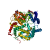 7c8kC 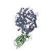 6lzgS S: Starting model for refinement C: citing same article ( |
|---|---|
| Similar structure data |


-Links
 PDBj
PDBj


- Assembly
| Deposited unit | 
| ||||||||||||
|---|---|---|---|---|---|---|---|---|---|---|---|---|---|
| 1 |
| ||||||||||||
| Unit cell |
|
-Components
| #1: Protein | Mass: 82350.930 Da / Num. of mol.: 1 Source method: isolated from a genetically manipulated source Source: (gene. exp.) Rhinolophus macrotis (Big-eared Horseshoe Bat)Gene: ACE2 / Production host:  References: UniProt: E2DHI3, Hydrolases; Acting on peptide bonds (peptidases) |
|---|---|
| #2: Protein | Mass: 21873.496 Da / Num. of mol.: 1 / Fragment: UNP residues 333-527 Source method: isolated from a genetically manipulated source Source: (gene. exp.) Severe acute respiratory syndrome coronavirus 2Gene: S, 2 Production host: Insect cell expression vector pTIE1 (others) References: UniProt: P0DTC2 |
| #3: Chemical | ChemComp-ZN /   Mass: 65.409 Da / Num. of mol.: 1 / Source method: obtained synthetically / Formula: Zn / Feature type: SUBJECT OF INVESTIGATION Mass: 65.409 Da / Num. of mol.: 1 / Source method: obtained synthetically / Formula: Zn / Feature type: SUBJECT OF INVESTIGATION |
| Has ligand of interest | Y |
| Has protein modification | Y |
-Experimental details
-Experiment
| Experiment | Method: X-RAY DIFFRACTION / Number of used crystals: 1 |
|---|
- Sample preparation
| Crystal | Density Matthews: 3.38 Å3/Da / Density % sol: 63.62 % |
|---|---|
| Crystal grow | Temperature: 291 K / Method: vapor diffusion, sitting drop Details: 0.1 M Succinic acid pH 7.0, 0.1 M BICINE pH 8.5, 30% v/v Polyethylene glycol monomethyl ether 550 |
-Data collection
| Diffraction | Mean temperature: 100 K / Serial crystal experiment: N |
|---|---|
| Diffraction source | Source: SYNCHROTRON / Site: SSRF / Beamline: BL19U1 / Wavelength: 0.97853 Å |
| Detector | Type: SDMS / Detector: CMOS / Date: May 10, 2020 |
| Radiation | Protocol: SINGLE WAVELENGTH / Monochromatic (M) / Laue (L): M / Scattering type: x-ray |
| Radiation wavelength | Wavelength: 0.97853 Å / Relative weight: 1 |
| Reflection | Resolution: 3.18→48.1 Å / Num. obs: 22038 / % possible obs: 99.9 % / Redundancy: 13.4 % / Biso Wilson estimate: 65.43 Å2 / Rmerge(I) obs: 0.163 / Net I/σ(I): 15.6 |
| Reflection shell | Resolution: 3.18→48.1 Å / Rmerge(I) obs: 1.79 |
- Processing
| Software |
| |||||||||||||||||||||||||||||||||||||||||||||||||||||||||||||||||||||||||||
|---|---|---|---|---|---|---|---|---|---|---|---|---|---|---|---|---|---|---|---|---|---|---|---|---|---|---|---|---|---|---|---|---|---|---|---|---|---|---|---|---|---|---|---|---|---|---|---|---|---|---|---|---|---|---|---|---|---|---|---|---|---|---|---|---|---|---|---|---|---|---|---|---|---|---|---|---|
| Refinement | Method to determine structure: MOLECULAR REPLACEMENT Starting model: 6LZG Resolution: 3.18→48.1 Å / SU ML: 0.3609 / Cross valid method: FREE R-VALUE / σ(F): 1.35 / Phase error: 27.2328 Stereochemistry target values: GeoStd + Monomer Library + CDL v1.2
| |||||||||||||||||||||||||||||||||||||||||||||||||||||||||||||||||||||||||||
| Solvent computation | Shrinkage radii: 0.9 Å / VDW probe radii: 1.11 Å / Solvent model: FLAT BULK SOLVENT MODEL | |||||||||||||||||||||||||||||||||||||||||||||||||||||||||||||||||||||||||||
| Displacement parameters | Biso mean: 62.21 Å2 | |||||||||||||||||||||||||||||||||||||||||||||||||||||||||||||||||||||||||||
| Refinement step | Cycle: LAST / Resolution: 3.18→48.1 Å
| |||||||||||||||||||||||||||||||||||||||||||||||||||||||||||||||||||||||||||
| Refine LS restraints |
| |||||||||||||||||||||||||||||||||||||||||||||||||||||||||||||||||||||||||||
| LS refinement shell |
| |||||||||||||||||||||||||||||||||||||||||||||||||||||||||||||||||||||||||||
| Refinement TLS params. | Method: refined / Refine-ID: X-RAY DIFFRACTION
| |||||||||||||||||||||||||||||||||||||||||||||||||||||||||||||||||||||||||||
| Refinement TLS group |
|
