 Movie
Movie Controller


+ Open data

- Basic information
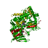
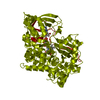
| Entry | Database: PDB / ID: 7c4m | |||||||||
|---|---|---|---|---|---|---|---|---|---|---|
| Title | Ancestral L-amino acid oxidase (AncLAAO-N5) L-Trp binding form | |||||||||
 Components Components | Ancestral L-amino acid oxidase | |||||||||
 Keywords Keywords | OXIDOREDUCTASE / L-amino acid oxidase | |||||||||
| Function / homology | FLAVIN-ADENINE DINUCLEOTIDE / TRYPTOPHAN Function and homology information Function and homology information | |||||||||
| Biological species | synthetic construct (others) | |||||||||
| Method |  X-RAY DIFFRACTION / SYNCHROTRON / SAD / Resolution: 2.4 Å X-RAY DIFFRACTION / SYNCHROTRON / SAD / Resolution: 2.4 Å | |||||||||
 Authors Authors | Nakano, S. / Minamino, Y. / Karasuda, H. / Ito, S. | |||||||||
| Funding support |  Japan, 2items Japan, 2items
| |||||||||
 Citation Citation | Journal: Commun Chem / Year: 2020 Title: Ancestral L-amino acid oxidases for deracemization and stereoinversion of amino acids Authors: Nakano, S. / Kozuka, K. / Minamino, Y. / Karasuda, H. / Hasebe, F. / Ito, S. | |||||||||
| History |
|
- Structure visualization
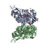
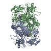
| Structure viewer | Molecule: MolmilJmol/JSmol |
|---|
- Downloads & links
-Download
| PDBx/mmCIF format | 7c4m.cif.gz | 173 KB | Display | PDBx/mmCIF format |
|---|---|---|---|---|
| PDB format | pdb7c4m.ent.gz | 109.4 KB | Display | PDB format |
| PDBx/mmJSON format | 7c4m.json.gz | Tree view | PDBx/mmJSON format | |
| Others |  Other downloads Other downloads |
-Validation report
| Arichive directory | https://data.pdbj.org/pub/pdb/validation_reports/c4/7c4mftp://data.pdbj.org/pub/pdb/validation_reports/c4/7c4m | HTTPS FTP |
|---|
-Related structure data


-Links
 PDBj
PDBj
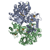
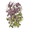
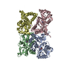
- Assembly
| Deposited unit | 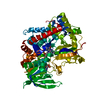
| ||||||||||||
|---|---|---|---|---|---|---|---|---|---|---|---|---|---|
| 1 | 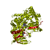
| ||||||||||||
| Unit cell |
|
-Components
| #1: Protein | Mass: 74963.977 Da / Num. of mol.: 1 / Source method: obtained synthetically / Source: (synth.) synthetic construct (others) |
|---|---|
| #2: Chemical | ChemComp-FAD /   Mass: 785.550 Da / Num. of mol.: 1 / Source method: obtained synthetically / Formula: C27H33N9O15P2 / Feature type: SUBJECT OF INVESTIGATION / Comment: FAD*YM Mass: 785.550 Da / Num. of mol.: 1 / Source method: obtained synthetically / Formula: C27H33N9O15P2 / Feature type: SUBJECT OF INVESTIGATION / Comment: FAD*YM |
| #3: Chemical | ChemComp-TRP /   Type: L-peptide linking / Mass: 204.225 Da / Num. of mol.: 1 / Source method: obtained synthetically / Formula: C11H12N2O2 Type: L-peptide linking / Mass: 204.225 Da / Num. of mol.: 1 / Source method: obtained synthetically / Formula: C11H12N2O2 |
| #4: Water | ChemComp-HOH /  Mass: 18.015 Da / Num. of mol.: 215 / Source method: isolated from a natural source / Formula: H2O Mass: 18.015 Da / Num. of mol.: 215 / Source method: isolated from a natural source / Formula: H2O |
| Has ligand of interest | Y |
-Experimental details
-Experiment
| Experiment | Method: X-RAY DIFFRACTION / Number of used crystals: 1 |
|---|
- Sample preparation
| Crystal | Density Matthews: 2.81 Å3/Da / Density % sol: 56.17 % |
|---|---|
| Crystal grow | Temperature: 295 K / Method: vapor diffusion, sitting drop / pH: 3.5 / Details: 1.0M NaCl, 0.1M citric acid pH 3.5 |
-Data collection
| Diffraction | Mean temperature: 100 K / Serial crystal experiment: N |
|---|---|
| Diffraction source | Source: SYNCHROTRON / Site: Photon Factory / Beamline: BL-5A / Wavelength: 1 Å |
| Detector | Type: DECTRIS PILATUS3 2M / Detector: PIXEL / Date: Dec 6, 2019 |
| Radiation | Protocol: SINGLE WAVELENGTH / Monochromatic (M) / Laue (L): M / Scattering type: x-ray |
| Radiation wavelength | Wavelength: 1 Å / Relative weight: 1 |
| Reflection | Resolution: 2.4→47.93 Å / Num. obs: 33554 / % possible obs: 100 % / Redundancy: 26.8 % / Biso Wilson estimate: 35.47 Å2 / CC1/2: 1 / Rmerge(I) obs: 0.107 / Net I/σ(I): 30.8 |
| Reflection shell | Resolution: 2.4→2.53 Å / Rmerge(I) obs: 0.596 / Num. unique obs: 4819 / CC1/2: 0.966 |
- Processing
| Software |
| |||||||||||||||||||||||||||||||||||||||||||||||||||||||||||||||||||||||||||||||||||||||||||
|---|---|---|---|---|---|---|---|---|---|---|---|---|---|---|---|---|---|---|---|---|---|---|---|---|---|---|---|---|---|---|---|---|---|---|---|---|---|---|---|---|---|---|---|---|---|---|---|---|---|---|---|---|---|---|---|---|---|---|---|---|---|---|---|---|---|---|---|---|---|---|---|---|---|---|---|---|---|---|---|---|---|---|---|---|---|---|---|---|---|---|---|---|
| Refinement | Method to determine structure: SAD / Resolution: 2.4→47.93 Å / SU ML: 0.3172 / Cross valid method: FREE R-VALUE / σ(F): 1.37 / Phase error: 25.2419 / Stereochemistry target values: CDL v1.2
| |||||||||||||||||||||||||||||||||||||||||||||||||||||||||||||||||||||||||||||||||||||||||||
| Solvent computation | Shrinkage radii: 0.9 Å / VDW probe radii: 1.11 Å / Solvent model: FLAT BULK SOLVENT MODEL | |||||||||||||||||||||||||||||||||||||||||||||||||||||||||||||||||||||||||||||||||||||||||||
| Displacement parameters | Biso mean: 37.95 Å2 | |||||||||||||||||||||||||||||||||||||||||||||||||||||||||||||||||||||||||||||||||||||||||||
| Refinement step | Cycle: LAST / Resolution: 2.4→47.93 Å
| |||||||||||||||||||||||||||||||||||||||||||||||||||||||||||||||||||||||||||||||||||||||||||
| Refine LS restraints |
| |||||||||||||||||||||||||||||||||||||||||||||||||||||||||||||||||||||||||||||||||||||||||||
| LS refinement shell |
|
