 Movie
Movie Controller

[English] 日本語
 Yorodumi
- PDB-2oov: Crystal Structure of Hansenula polymorpha amine oxidase to 1.7 An... -
+ Open data

- Basic information
| Entry | Database: PDB / ID: 2oov | ||||||
|---|---|---|---|---|---|---|---|
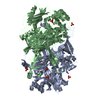
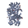
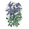
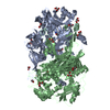
| Title | Crystal Structure of Hansenula polymorpha amine oxidase to 1.7 Angstroms | ||||||
 Components Components | (Peroxisomal copper amine ...) x 2 | ||||||
 Keywords Keywords | OXIDOREDUCTASE / protein-derived cofactor / beta-sandwich / TPQ | ||||||
| Function / homology |  Function and homology information Function and homology informationprimary-amine oxidase / primary methylamine oxidase activity / amine metabolic process / quinone binding / peroxisome / copper ion binding Similarity search - Function | ||||||
| Biological species |  Pichia angusta (fungus) Pichia angusta (fungus) | ||||||
| Method |  X-RAY DIFFRACTION / SYNCHROTRON / MOLECULAR REPLACEMENT / Resolution: 1.7 Å X-RAY DIFFRACTION / SYNCHROTRON / MOLECULAR REPLACEMENT / Resolution: 1.7 Å | ||||||
 Authors Authors | Johnson, B.J. / Wilmot, C.M. | ||||||
 Citation Citation | Journal: J.Biol.Chem. / Year: 2007 Title: Exploring molecular oxygen pathways in Hansenula polymorpha copper-containing amine oxidase Authors: Johnson, B.J. / Cohen, J. / Welford, R.W. / Pearson, A.R. / Schulten, K. / Klinman, J.P. / Wilmot, C.M. | ||||||
| History |
| ||||||
| Remark 999 | SEQUENCE The author states that there is radiation damage at MET634 in the protein that is an ... SEQUENCE The author states that there is radiation damage at MET634 in the protein that is an oxidation clearly visible in chains A and B, but not in chains C,D,E, or F. The protein that went into the crystallization had an unmodified MET at position 634. |
- Structure visualization
| Structure viewer | Molecule: MolmilJmol/JSmol |
|---|
- Downloads & links
-Download
| PDBx/mmCIF format | 2oov.cif.gz | 890.5 KB | Display | PDBx/mmCIF format |
|---|---|---|---|---|
| PDB format | pdb2oov.ent.gz | 724.3 KB | Display | PDB format |
| PDBx/mmJSON format | 2oov.json.gz | Tree view | PDBx/mmJSON format | |
| Others |  Other downloads Other downloads |
-Validation report
| Arichive directory | https://data.pdbj.org/pub/pdb/validation_reports/oo/2oovftp://data.pdbj.org/pub/pdb/validation_reports/oo/2oov | HTTPS FTP |
|---|
-Related structure data
| Related structure data |  2oqeC  1a2vS S: Starting model for refinement C: citing same article ( |
|---|---|
| Similar structure data |


-Links
 PDBj
PDBj- Assembly
| Deposited unit | 
| ||||||||
|---|---|---|---|---|---|---|---|---|---|
| 1 | 
| ||||||||
| 2 | 
| ||||||||
| 3 | 
| ||||||||
| 4 |
| ||||||||
| Unit cell |
| ||||||||
| Details | The biological assembly is a dimer. The asymmetric unit contains three dimers. |
-Components
-Peroxisomal copper amine ... , 2 types, 6 molecules ABCDEF
| #1: Protein | Mass: 74077.438 Da / Num. of mol.: 2 / Fragment: Residues 13-672 Source method: isolated from a genetically manipulated source Source: (gene. exp.) Pichia angusta (fungus) / Gene: AMO / Plasmid: pDB20 / Production host:  #2: Protein | Mass: 74061.438 Da / Num. of mol.: 4 / Fragment: Residues 13-672 Source method: isolated from a genetically manipulated source Source: (gene. exp.) Pichia angusta (fungus) / Gene: AMO / Plasmid: pDB20 / Production host: |
|---|
-Non-polymers , 4 types, 5091 molecules 


| #3: Chemical | ChemComp-CU /  Mass: 63.546 Da / Num. of mol.: 6 / Source method: obtained synthetically / Formula: Cu Mass: 63.546 Da / Num. of mol.: 6 / Source method: obtained synthetically / Formula: Cu#4: Chemical | ChemComp-GOL /  Mass: 92.094 Da / Num. of mol.: 28 / Source method: obtained synthetically / Formula: C3H8O3 Mass: 92.094 Da / Num. of mol.: 28 / Source method: obtained synthetically / Formula: C3H8O3#5: Chemical |  Mass: 94.971 Da / Num. of mol.: 2 / Source method: obtained synthetically / Formula: PO4 Mass: 94.971 Da / Num. of mol.: 2 / Source method: obtained synthetically / Formula: PO4#6: Water | ChemComp-HOH / | Mass: 18.015 Da / Num. of mol.: 5055 / Source method: isolated from a natural source / Formula: H2O |
|---|
-Experimental details
-Experiment
| Experiment | Method: X-RAY DIFFRACTION / Number of used crystals: 1 |
|---|
- Sample preparation
| Crystal | Density Matthews: 2.72 Å3/Da / Density % sol: 54.83 % |
|---|---|
| Crystal grow | Temperature: 298 K / Method: vapor diffusion, hanging drop / pH: 6 Details: 8% PEG 8000, 0.3M potassium phosphate, pH 6.0, VAPOR DIFFUSION, HANGING DROP, temperature 298.0K |
-Data collection
| Diffraction | Mean temperature: 100 K |
|---|---|
| Diffraction source | Source: SYNCHROTRON / Site: APS  / Beamline: 19-ID / Wavelength: 0.979 Å / Beamline: 19-ID / Wavelength: 0.979 Å |
| Detector | Type: ADSC QUANTUM 315 / Detector: CCD / Date: Jul 30, 2005 |
| Radiation | Monochromator: Rosenbaum-Rock high-resolution double-crystal Protocol: SINGLE WAVELENGTH / Monochromatic (M) / Laue (L): M / Scattering type: x-ray |
| Radiation wavelength | Wavelength: 0.979 Å / Relative weight: 1 |
| Reflection | Resolution: 1.7→50 Å / Num. obs: 437335 / % possible obs: 85.1 % / Redundancy: 4.6 % / Rmerge(I) obs: 0.085 / Χ2: 0.96 / Net I/σ(I): 8.4 |
| Reflection shell | Resolution: 1.7→1.76 Å / Redundancy: 2.8 % / Rmerge(I) obs: 0.55 / Num. unique all: 18134 / Χ2: 0.706 / % possible all: 35.3 |
-Phasing
| Phasing MR | Rfactor: 0.301 / Cor.coef. Fo:Fc: 0.736
|
|---|
- Processing
| Software |
| ||||||||||||||||||||||||||||||||||||||||||||||||||||||||||||||||||||||||||||||||||||||||||
|---|---|---|---|---|---|---|---|---|---|---|---|---|---|---|---|---|---|---|---|---|---|---|---|---|---|---|---|---|---|---|---|---|---|---|---|---|---|---|---|---|---|---|---|---|---|---|---|---|---|---|---|---|---|---|---|---|---|---|---|---|---|---|---|---|---|---|---|---|---|---|---|---|---|---|---|---|---|---|---|---|---|---|---|---|---|---|---|---|---|---|---|
| Refinement | Method to determine structure: MOLECULAR REPLACEMENT Starting model: PDB entry 1A2V Resolution: 1.7→37.96 Å / Cor.coef. Fo:Fc: 0.968 / Cor.coef. Fo:Fc free: 0.955 / SU B: 1.725 / SU ML: 0.057 / Cross valid method: THROUGHOUT / σ(F): 0 / ESU R: 0.099 / ESU R Free: 0.097 Stereochemistry target values: MAXIMUM LIKELIHOOD WITH PHASES Details: HYDROGENS HAVE BEEN ADDED IN THE RIDING POSITIONS. An occupancy greater than 1.0 of atom O4 on residue 405 was used to correctly model a water that is at this position when the cofactor is ...Details: HYDROGENS HAVE BEEN ADDED IN THE RIDING POSITIONS. An occupancy greater than 1.0 of atom O4 on residue 405 was used to correctly model a water that is at this position when the cofactor is in the alternate conformer. This was required because refmac did not refine a 0.5 occupancy water and 0.5 occupancy O4 in an identical position correctly. This problem will exist at the O4 of residue 405 in chains C,D,E and F.
| ||||||||||||||||||||||||||||||||||||||||||||||||||||||||||||||||||||||||||||||||||||||||||
| Solvent computation | Ion probe radii: 0.8 Å / Shrinkage radii: 0.8 Å / VDW probe radii: 1.4 Å / Solvent model: BABINET MODEL WITH MASK | ||||||||||||||||||||||||||||||||||||||||||||||||||||||||||||||||||||||||||||||||||||||||||
| Displacement parameters | Biso mean: 22.284 Å2
| ||||||||||||||||||||||||||||||||||||||||||||||||||||||||||||||||||||||||||||||||||||||||||
| Refinement step | Cycle: LAST / Resolution: 1.7→37.96 Å
| ||||||||||||||||||||||||||||||||||||||||||||||||||||||||||||||||||||||||||||||||||||||||||
| Refine LS restraints |
| ||||||||||||||||||||||||||||||||||||||||||||||||||||||||||||||||||||||||||||||||||||||||||
| LS refinement shell | Resolution: 1.7→1.746 Å / Total num. of bins used: 20
|
