 Movie
Movie Controller

[English] 日本語
 Yorodumi
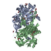
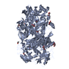
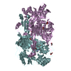
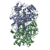
- PDB-1ekm: CRYSTAL STRUCTURE AT 2.5 A RESOLUTION OF ZINC-SUBSTITUTED COPPER ... -
+ Open data

- Basic information
| Entry | Database: PDB / ID: 1ekm | ||||||
|---|---|---|---|---|---|---|---|
| Title | CRYSTAL STRUCTURE AT 2.5 A RESOLUTION OF ZINC-SUBSTITUTED COPPER AMINE OXIDASE OF HANSENULA POLYMORPHA EXPRESSED IN ESCHERICHIA COLI | ||||||
 Components Components | COPPER AMINE OXIDASE | ||||||
 Keywords Keywords | OXIDOREDUCTASE / amine oxidase / quinoprotein | ||||||
| Function / homology |  Function and homology information Function and homology informationprimary-amine oxidase / primary methylamine oxidase activity / amine metabolic process / quinone binding / peroxisome / copper ion binding Similarity search - Function | ||||||
| Biological species |  Pichia angusta (fungus) Pichia angusta (fungus) | ||||||
| Method |  X-RAY DIFFRACTION / Resolution: 2.5 Å X-RAY DIFFRACTION / Resolution: 2.5 Å | ||||||
 Authors Authors | Chen, Z. / Schwartz, B. / Williams, N.K. / Li, R. / Klinman, J.P. / Mathews, F.S. | ||||||
 Citation Citation | Journal: Biochemistry / Year: 2000 Title: Crystal structure at 2.5 A resolution of zinc-substituted copper amine oxidase of Hansenula polymorpha expressed in Escherichia coli. Authors: Chen, Z. / Schwartz, B. / Williams, N.K. / Li, R. / Klinman, J.P. / Mathews, F.S. #1: Journal: Structure / Year: 1998Title: Copper Amine Oxidase from Hansenula polymorpha: the Crystal Structure Determined at 2.4 A Resolution Reveals the Active Conformation Authors: Li, R. / Klinman, J.P. / Mathews, F.S. | ||||||
| History |
|
- Structure visualization
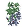
| Structure viewer | Molecule: MolmilJmol/JSmol |
|---|
- Downloads & links
-Download
| PDBx/mmCIF format | 1ekm.cif.gz | 420 KB | Display | PDBx/mmCIF format |
|---|---|---|---|---|
| PDB format | pdb1ekm.ent.gz | 340 KB | Display | PDB format |
| PDBx/mmJSON format | 1ekm.json.gz | Tree view | PDBx/mmJSON format | |
| Others |  Other downloads Other downloads |
-Validation report
| Arichive directory | https://data.pdbj.org/pub/pdb/validation_reports/ek/1ekmftp://data.pdbj.org/pub/pdb/validation_reports/ek/1ekm | HTTPS FTP |
|---|
-Related structure data
| Related structure data | |
|---|---|
| Similar structure data |


-Links
 PDBj
PDBj- Assembly
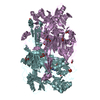
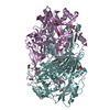
| Deposited unit | 
| ||||||||||||
|---|---|---|---|---|---|---|---|---|---|---|---|---|---|
| 1 | 
| ||||||||||||
| 2 | 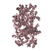
| ||||||||||||
| 3 | 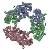
| ||||||||||||
| Unit cell |
| ||||||||||||
| Noncrystallographic symmetry (NCS) | NCS oper:
| ||||||||||||
| Details | Molecule is homodimer in solution. The biological unit is a dimer. Three dimers form a crystallographic hexamer which is non-biological. The asymmetric unit contains half of a hexamer, chains ABC. Chains B,C were not treated as independent during refinement but were generated by strict non-crystallographic symmetry. |
-Components
| #1: Protein | Mass: 73731.141 Da / Num. of mol.: 3 Source method: isolated from a genetically manipulated source Details: ZINC-SUBSTITUTED / Source: (gene. exp.) Pichia angusta (fungus) / Plasmid: PET11A / Production host:  #2: Chemical |   Mass: 65.409 Da / Num. of mol.: 3 / Source method: obtained synthetically / Formula: Zn Mass: 65.409 Da / Num. of mol.: 3 / Source method: obtained synthetically / Formula: Zn#3: Water | ChemComp-HOH / |  Mass: 18.015 Da / Num. of mol.: 1476 / Source method: isolated from a natural source / Formula: H2O Mass: 18.015 Da / Num. of mol.: 1476 / Source method: isolated from a natural source / Formula: H2OHas protein modification | Y | |
|---|
-Experimental details
-Experiment
| Experiment | Method: X-RAY DIFFRACTION / Number of used crystals: 1 |
|---|
- Sample preparation
| Crystal | Density Matthews: 2.7 Å3/Da / Density % sol: 54.39 % | ||||||||||||||||||||
|---|---|---|---|---|---|---|---|---|---|---|---|---|---|---|---|---|---|---|---|---|---|
| Crystal grow | Temperature: 295 K / Method: vapor diffusion, hanging drop Details: PEG 8000, potassium phosphate, VAPOR DIFFUSION, HANGING DROP, temperature 295.0K | ||||||||||||||||||||
| Crystal | *PLUS Density % sol: 52 % | ||||||||||||||||||||
| Crystal grow | *PLUS PH range low: 7.5 / PH range high: 6.2 | ||||||||||||||||||||
| Components of the solutions | *PLUS
|
-Data collection
| Diffraction | Mean temperature: 110 K |
|---|---|
| Diffraction source | Source: ROTATING ANODE / Type: RIGAKU RU200 / Wavelength: 1.5418 |
| Detector | Type: RIGAKU RAXIS / Detector: IMAGE PLATE / Date: Sep 25, 1998 |
| Radiation | Protocol: SINGLE WAVELENGTH / Monochromatic (M) / Laue (L): M / Scattering type: x-ray |
| Radiation wavelength | Wavelength: 1.5418 Å / Relative weight: 1 |
| Reflection | Resolution: 2.5→500 Å / Num. all: 65122 / Num. obs: 64796 / % possible obs: 99.5 % / Observed criterion σ(F): 0 / Observed criterion σ(I): 0 / Redundancy: 3.5 % / Biso Wilson estimate: 27.4 Å2 / Rmerge(I) obs: 0.087 / Net I/σ(I): 11.8 |
| Reflection shell | Resolution: 2.5→2.6 Å / Redundancy: 2.46 % / Rmerge(I) obs: 0.185 / Num. unique all: 3689 / % possible all: 40.7 |
| Reflection | *PLUS % possible obs: 77.4 % / Num. measured all: 226364 |
| Reflection shell | *PLUS % possible obs: 40.7 % / Mean I/σ(I) obs: 3 |
- Processing
| Software |
| |||||||||||||||||||||||||
|---|---|---|---|---|---|---|---|---|---|---|---|---|---|---|---|---|---|---|---|---|---|---|---|---|---|---|
| Refinement | Resolution: 2.5→500 Å / σ(F): 0 / σ(I): 0 / Stereochemistry target values: ENGH & HUBER / Details: USED STRICT NCS CONSTRAINTS
| |||||||||||||||||||||||||
| Refinement step | Cycle: LAST / Resolution: 2.5→500 Å
| |||||||||||||||||||||||||
| Refine LS restraints |
| |||||||||||||||||||||||||
| Software | *PLUS Name: CNS / Classification: refinement | |||||||||||||||||||||||||
| Refinement | *PLUS % reflection Rfree: 10 % | |||||||||||||||||||||||||
| Solvent computation | *PLUS | |||||||||||||||||||||||||
| Displacement parameters | *PLUS |
