 Movie
Movie Controller

[English] 日本語
 Yorodumi
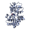
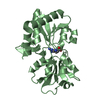
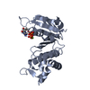
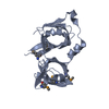
- PDB-7bzk: Crystal structure of ferredoxin: thioredoxin reductase and thiore... -
+ Open data

- Basic information
| Entry | Database: PDB / ID: 7bzk | ||||||
|---|---|---|---|---|---|---|---|
| Title | Crystal structure of ferredoxin: thioredoxin reductase and thioredoxin y1 complex | ||||||
 Components Components |
| ||||||
 Keywords Keywords | ELECTRON TRANSPORT / FTR / Trx y1 | ||||||
| Function / homology |  Function and homology information Function and homology informationferredoxin-thioredoxin reductase activity / ferredoxin:thioredoxin reductase / oxidoreductase activity, acting on iron-sulfur proteins as donors / positive regulation of catalytic activity / chloroplast envelope / chloroplast stroma / protein-disulfide reductase activity / photosynthesis / chloroplast / enzyme activator activity ...ferredoxin-thioredoxin reductase activity / ferredoxin:thioredoxin reductase / oxidoreductase activity, acting on iron-sulfur proteins as donors / positive regulation of catalytic activity / chloroplast envelope / chloroplast stroma / protein-disulfide reductase activity / photosynthesis / chloroplast / enzyme activator activity / 4 iron, 4 sulfur cluster binding / electron transfer activity / metal ion binding Similarity search - Function | ||||||
| Biological species |  | ||||||
| Method |  X-RAY DIFFRACTION / SYNCHROTRON / MOLECULAR REPLACEMENT / Resolution: 1.5935 Å X-RAY DIFFRACTION / SYNCHROTRON / MOLECULAR REPLACEMENT / Resolution: 1.5935 Å | ||||||
 Authors Authors | Kurisu, G. / Juniar, L. / Tanaka, H. | ||||||
| Funding support |  Japan, 1items Japan, 1items
| ||||||
 Citation Citation | Journal: Protein Sci. / Year: 2020 Title: Structural basis for thioredoxin isoform-based fine-tuning of ferredoxin-thioredoxin reductase activity. Authors: Juniar, L. / Tanaka, H. / Yoshida, K. / Hisabori, T. / Kurisu, G. | ||||||
| History |
|
- Structure visualization
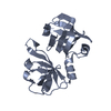
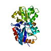
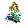
| Structure viewer | Molecule: MolmilJmol/JSmol |
|---|
- Downloads & links
-Download
| PDBx/mmCIF format | 7bzk.cif.gz | 59.4 KB | Display | PDBx/mmCIF format |
|---|---|---|---|---|
| PDB format | pdb7bzk.ent.gz | 41.7 KB | Display | PDB format |
| PDBx/mmJSON format | 7bzk.json.gz | Tree view | PDBx/mmJSON format | |
| Others |  Other downloads Other downloads |
-Validation report
| Arichive directory | https://data.pdbj.org/pub/pdb/validation_reports/bz/7bzkftp://data.pdbj.org/pub/pdb/validation_reports/bz/7bzk | HTTPS FTP |
|---|
-Related structure data
| Related structure data | 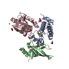 7c2bC  7c3fC  7c65C  2pu9S S: Starting model for refinement C: citing same article ( |
|---|---|
| Similar structure data |


-Links
 PDBj
PDBj

- Assembly
| Deposited unit | 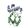
| ||||||||
|---|---|---|---|---|---|---|---|---|---|
| 1 |
| ||||||||
| Unit cell |
|
-Components
| #1: Protein | Mass: 12995.734 Da / Num. of mol.: 1 Source method: isolated from a genetically manipulated source Source: (gene. exp.)  References: UniProt: Q9SJ89, ferredoxin:thioredoxin reductase |
|---|---|
| #2: Protein | Mass: 12601.493 Da / Num. of mol.: 1 / Mutation: C34S Source method: isolated from a genetically manipulated source Source: (gene. exp.) |
| #3: Chemical | ChemComp-SF4 /   Mass: 351.640 Da / Num. of mol.: 1 / Source method: obtained synthetically / Formula: Fe4S4 / Feature type: SUBJECT OF INVESTIGATION Mass: 351.640 Da / Num. of mol.: 1 / Source method: obtained synthetically / Formula: Fe4S4 / Feature type: SUBJECT OF INVESTIGATION |
| #4: Water | ChemComp-HOH /  Mass: 18.015 Da / Num. of mol.: 108 / Source method: isolated from a natural source / Formula: H2O Mass: 18.015 Da / Num. of mol.: 108 / Source method: isolated from a natural source / Formula: H2O |
| Has ligand of interest | Y |
| Has protein modification | Y |
-Experimental details
-Experiment
| Experiment | Method: X-RAY DIFFRACTION / Number of used crystals: 1 |
|---|
- Sample preparation
| Crystal | Density Matthews: 2 Å3/Da / Density % sol: 38.58 % |
|---|---|
| Crystal grow | Temperature: 293.15 K / Method: vapor diffusion, hanging drop / pH: 3.5 Details: 100 mM Magnesium Citric acid pH 3.5 5% 2-propanol 5-9% PEG 3,350 PH range: 3.5-7.5 |
-Data collection
| Diffraction | Mean temperature: 100 K / Serial crystal experiment: N |
|---|---|
| Diffraction source | Source: SYNCHROTRON / Site: SPring-8 / Beamline: BL44XU / Wavelength: 0.9 Å |
| Detector | Type: DECTRIS EIGER X 16M / Detector: PIXEL / Date: Feb 4, 2020 |
| Radiation | Protocol: SINGLE WAVELENGTH / Monochromatic (M) / Laue (L): M / Scattering type: x-ray |
| Radiation wavelength | Wavelength: 0.9 Å / Relative weight: 1 |
| Reflection | Resolution: 1.5935→43.0845 Å / Num. obs: 27925 / % possible obs: 99.89 % / Redundancy: 6.6 % / Biso Wilson estimate: 22.87 Å2 / CC1/2: 0.995 / CC star: 0.999 / Rmerge(I) obs: 0.1197 / Rpim(I) all: 0.05048 / Rrim(I) all: 0.1301 / Net I/σ(I): 10.4 |
| Reflection shell | Resolution: 1.5935→1.651 Å / Redundancy: 6.2 % / Rmerge(I) obs: 0.7606 / Num. unique obs: 2737 / CC1/2: 0.861 / CC star: 0.962 / Rpim(I) all: 0.3309 / Rrim(I) all: 0.8309 / % possible all: 99.56 |
- Processing
| Software |
| ||||||||||||||||||||||||||||||||||||||||||||||||||||||||||||||||||
|---|---|---|---|---|---|---|---|---|---|---|---|---|---|---|---|---|---|---|---|---|---|---|---|---|---|---|---|---|---|---|---|---|---|---|---|---|---|---|---|---|---|---|---|---|---|---|---|---|---|---|---|---|---|---|---|---|---|---|---|---|---|---|---|---|---|---|---|
| Refinement | Method to determine structure: MOLECULAR REPLACEMENT Starting model: 2PU9 Resolution: 1.5935→43.0845 Å / SU ML: 0.17 / Cross valid method: THROUGHOUT / σ(F): 1.37 / Phase error: 19.54
| ||||||||||||||||||||||||||||||||||||||||||||||||||||||||||||||||||
| Solvent computation | Shrinkage radii: 0.9 Å / VDW probe radii: 1.11 Å | ||||||||||||||||||||||||||||||||||||||||||||||||||||||||||||||||||
| Displacement parameters | Biso max: 87.41 Å2 / Biso mean: 22.45 Å2 / Biso min: 8.97 Å2 | ||||||||||||||||||||||||||||||||||||||||||||||||||||||||||||||||||
| Refinement step | Cycle: final / Resolution: 1.5935→43.0845 Å
| ||||||||||||||||||||||||||||||||||||||||||||||||||||||||||||||||||
| LS refinement shell | Refine-ID: X-RAY DIFFRACTION / Rfactor Rfree error: 0
|
