 Movie
Movie Controller

[English] 日本語
 Yorodumi
- PDB-7bzg: Structure of Bacillus subtilis HxlR, wild type in complex with fo... -
+ Open data

- Basic information
| Entry | Database: PDB / ID: 7bzg | |||||||||||||||
|---|---|---|---|---|---|---|---|---|---|---|---|---|---|---|---|---|
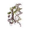
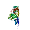
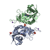
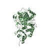
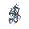
| Title | Structure of Bacillus subtilis HxlR, wild type in complex with formaldehyde and DNA | |||||||||||||||
 Components Components |
| |||||||||||||||
 Keywords Keywords | DNA BINDING PROTEIN / Transcriptional regulator / formaldehyde sensing / DNA BINDING PROTEIN-DNA complex | |||||||||||||||
| Function / homology |  Function and homology information Function and homology information | |||||||||||||||
| Biological species |  | |||||||||||||||
| Method |  X-RAY DIFFRACTION / SYNCHROTRON / MOLECULAR REPLACEMENT / Resolution: 2.9 Å X-RAY DIFFRACTION / SYNCHROTRON / MOLECULAR REPLACEMENT / Resolution: 2.9 Å | |||||||||||||||
 Authors Authors | Zhu, R. / Chen, P.R. | |||||||||||||||
| Funding support |  China, 4items China, 4items
| |||||||||||||||
 Citation Citation | Journal: Nat Commun / Year: 2021 Title: Genetically encoded formaldehyde sensors inspired by a protein intra-helical crosslinking reaction. Authors: Zhu, R. / Zhang, G. / Jing, M. / Han, Y. / Li, J. / Zhao, J. / Li, Y. / Chen, P.R. | |||||||||||||||
| History |
|
- Structure visualization
| Structure viewer | Molecule: MolmilJmol/JSmol |
|---|
- Downloads & links
-Download
| PDBx/mmCIF format | 7bzg.cif.gz | 223.5 KB | Display | PDBx/mmCIF format |
|---|---|---|---|---|
| PDB format | pdb7bzg.ent.gz | 171.3 KB | Display | PDB format |
| PDBx/mmJSON format | 7bzg.json.gz | Tree view | PDBx/mmJSON format | |
| Others |  Other downloads Other downloads |
-Validation report
| Arichive directory | https://data.pdbj.org/pub/pdb/validation_reports/bz/7bzgftp://data.pdbj.org/pub/pdb/validation_reports/bz/7bzg | HTTPS FTP |
|---|
-Related structure data
| Related structure data |  7bzdC  7bzeC 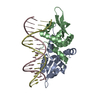 4hqeS C: citing same article ( S: Starting model for refinement |
|---|---|
| Similar structure data |


-Links
 PDBj
PDBj


- Assembly
| Deposited unit | 
| ||||||||
|---|---|---|---|---|---|---|---|---|---|
| 1 | 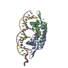
| ||||||||
| 2 | 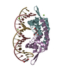
| ||||||||
| 3 | 
| ||||||||
| Unit cell |
|
-Components
-Protein / DNA chain , 2 types, 12 molecules ABEFIJCDGHKL
| #1: Protein | Mass: 14405.915 Da / Num. of mol.: 6 Source method: isolated from a genetically manipulated source Source: (gene. exp.) Strain: 168 / Gene: hxlR, BSU03470 / Production host: #2: DNA chain | Mass: 6133.979 Da / Num. of mol.: 6 / Source method: obtained synthetically Source: (synth.) |
|---|
-Non-polymers , 6 types, 198 molecules 


| #3: Chemical | ChemComp-FOR /  Mass: 30.026 Da / Num. of mol.: 6 Mass: 30.026 Da / Num. of mol.: 6Source method: isolated from a genetically manipulated source Formula: CH2O / Feature type: SUBJECT OF INVESTIGATION #4: Chemical | ChemComp-PGE /  Mass: 150.173 Da / Num. of mol.: 5 / Source method: obtained synthetically / Formula: C6H14O4 Mass: 150.173 Da / Num. of mol.: 5 / Source method: obtained synthetically / Formula: C6H14O4#5: Chemical | ChemComp-MG /  Mass: 24.305 Da / Num. of mol.: 7 / Source method: isolated from a natural source / Formula: Mg Mass: 24.305 Da / Num. of mol.: 7 / Source method: isolated from a natural source / Formula: Mg#6: Chemical | ChemComp-PE8 / |  Mass: 370.436 Da / Num. of mol.: 1 Mass: 370.436 Da / Num. of mol.: 1Source method: isolated from a genetically manipulated source Formula: C16H34O9 #7: Chemical | ChemComp-PEG / |  Mass: 106.120 Da / Num. of mol.: 1 / Source method: obtained synthetically / Formula: C4H10O3 Mass: 106.120 Da / Num. of mol.: 1 / Source method: obtained synthetically / Formula: C4H10O3#8: Water | ChemComp-HOH / | Mass: 18.015 Da / Num. of mol.: 178 / Source method: isolated from a natural source / Formula: H2O |
|---|
-Details
| Has ligand of interest | Y |
|---|---|
| Has protein modification | Y |
-Experimental details
-Experiment
| Experiment | Method: X-RAY DIFFRACTION / Number of used crystals: 1 |
|---|
- Sample preparation
| Crystal | Density Matthews: 3.93 Å3/Da / Density % sol: 70.95 % |
|---|---|
| Crystal grow | Temperature: 293 K / Method: vapor diffusion, sitting drop / Details: 0.1 MES pH = 6.4, 50 mM MgCl2, 25% v/v PEG MME 550 |
-Data collection
| Diffraction | Mean temperature: 100 K / Serial crystal experiment: N |
|---|---|
| Diffraction source | Source: SYNCHROTRON / Site: SSRF / Beamline: BL17U1 / Wavelength: 0.9793 Å |
| Detector | Type: ADSC QUANTUM 315r / Detector: CCD / Date: Oct 6, 2015 |
| Radiation | Protocol: SINGLE WAVELENGTH / Monochromatic (M) / Laue (L): M / Scattering type: x-ray |
| Radiation wavelength | Wavelength: 0.9793 Å / Relative weight: 1 |
| Reflection | Resolution: 2.9→50 Å / Num. obs: 42387 / % possible obs: 99.8 % / Redundancy: 3.8 % / Rmerge(I) obs: 0.095 / Net I/σ(I): 17.6 |
| Reflection shell | Resolution: 2.9→2.95 Å / Redundancy: 3.8 % / Rmerge(I) obs: 0.966 / Mean I/σ(I) obs: 2 / Num. unique obs: 2144 / % possible all: 100 |
- Processing
| Software |
| |||||||||||||||||||||||||||||||||||||||||||||||||||||||||||||||||||||||||||||||||||||||||||||||||||||||||
|---|---|---|---|---|---|---|---|---|---|---|---|---|---|---|---|---|---|---|---|---|---|---|---|---|---|---|---|---|---|---|---|---|---|---|---|---|---|---|---|---|---|---|---|---|---|---|---|---|---|---|---|---|---|---|---|---|---|---|---|---|---|---|---|---|---|---|---|---|---|---|---|---|---|---|---|---|---|---|---|---|---|---|---|---|---|---|---|---|---|---|---|---|---|---|---|---|---|---|---|---|---|---|---|---|---|---|
| Refinement | Method to determine structure: MOLECULAR REPLACEMENT Starting model: 4HQE Resolution: 2.9→33.409 Å / SU ML: 0.36 / Cross valid method: FREE R-VALUE / σ(F): 1.34 / Phase error: 23.45
| |||||||||||||||||||||||||||||||||||||||||||||||||||||||||||||||||||||||||||||||||||||||||||||||||||||||||
| Solvent computation | Shrinkage radii: 0.9 Å / VDW probe radii: 1.11 Å | |||||||||||||||||||||||||||||||||||||||||||||||||||||||||||||||||||||||||||||||||||||||||||||||||||||||||
| Refinement step | Cycle: LAST / Resolution: 2.9→33.409 Å
| |||||||||||||||||||||||||||||||||||||||||||||||||||||||||||||||||||||||||||||||||||||||||||||||||||||||||
| Refine LS restraints |
| |||||||||||||||||||||||||||||||||||||||||||||||||||||||||||||||||||||||||||||||||||||||||||||||||||||||||
| LS refinement shell |
|
