 Movie
Movie Controller


+ Open data

- Basic information
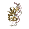
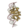
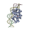
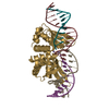
| Entry | Database: PDB / ID: 7bzd | |||||||||||||||
|---|---|---|---|---|---|---|---|---|---|---|---|---|---|---|---|---|
| Title | Structure of Bacillus subtilis HxlR, wild type | |||||||||||||||
 Components Components | HTH-type transcriptional activator HxlR | |||||||||||||||
 Keywords Keywords | DNA BINDING PROTEIN / Transcriptional regulator / formaldehyde sensing | |||||||||||||||
| Function / homology | Helix-turn-helix, HxlR type / HxlR-like helix-turn-helix / HxlR-type HTH domain profile. / Winged helix DNA-binding domain superfamily / Winged helix-like DNA-binding domain superfamily / DNA binding / HTH-type transcriptional activator HxlR Function and homology information Function and homology information | |||||||||||||||
| Biological species |  | |||||||||||||||
| Method |  X-RAY DIFFRACTION / SYNCHROTRON / MOLECULAR REPLACEMENT / Resolution: 2.612 Å X-RAY DIFFRACTION / SYNCHROTRON / MOLECULAR REPLACEMENT / Resolution: 2.612 Å | |||||||||||||||
 Authors Authors | Zhu, R. / Chen, P.R. | |||||||||||||||
| Funding support |  China, 4items China, 4items
| |||||||||||||||
 Citation Citation | Journal: Nat Commun / Year: 2021 Title: Genetically encoded formaldehyde sensors inspired by a protein intra-helical crosslinking reaction. Authors: Zhu, R. / Zhang, G. / Jing, M. / Han, Y. / Li, J. / Zhao, J. / Li, Y. / Chen, P.R. | |||||||||||||||
| History |
|
- Structure visualization
| Structure viewer | Molecule: MolmilJmol/JSmol |
|---|
- Downloads & links
-Download
| PDBx/mmCIF format | 7bzd.cif.gz | 36.5 KB | Display | PDBx/mmCIF format |
|---|---|---|---|---|
| PDB format | pdb7bzd.ent.gz | 22.9 KB | Display | PDB format |
| PDBx/mmJSON format | 7bzd.json.gz | Tree view | PDBx/mmJSON format | |
| Others |  Other downloads Other downloads |
-Validation report
| Arichive directory | https://data.pdbj.org/pub/pdb/validation_reports/bz/7bzdftp://data.pdbj.org/pub/pdb/validation_reports/bz/7bzd | HTTPS FTP |
|---|
-Related structure data
| Related structure data |  7bzeC 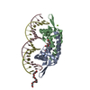 7bzgC  4a5nS S: Starting model for refinement C: citing same article ( |
|---|---|
| Similar structure data |


-Links
 PDBj
PDBj- Assembly
| Deposited unit | 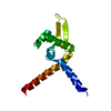
| ||||||||
|---|---|---|---|---|---|---|---|---|---|
| 1 | 
| ||||||||
| Unit cell |
|
-Components
| #1: Protein | Mass: 14405.915 Da / Num. of mol.: 1 Source method: isolated from a genetically manipulated source Source: (gene. exp.) Strain: 168 / Gene: hxlR, BSU03470 / Production host: |
|---|
-Experimental details
-Experiment
| Experiment | Method: X-RAY DIFFRACTION / Number of used crystals: 1 |
|---|
- Sample preparation
| Crystal | Density Matthews: 1.98 Å3/Da / Density % sol: 37.92 % |
|---|---|
| Crystal grow | Temperature: 293 K / Method: vapor diffusion, sitting drop Details: 0.1 M sodium acetate trihydrate pH = 4.6, 1.5 M ammonium chloride, 10 mM DTT |
-Data collection
| Diffraction | Mean temperature: 100 K / Serial crystal experiment: N |
|---|---|
| Diffraction source | Source: SYNCHROTRON / Site: SSRF / Beamline: BL17U1 / Wavelength: 0.97915 Å |
| Detector | Type: ADSC QUANTUM 315r / Detector: CCD / Date: Jan 14, 2015 |
| Radiation | Protocol: SINGLE WAVELENGTH / Monochromatic (M) / Laue (L): M / Scattering type: x-ray |
| Radiation wavelength | Wavelength: 0.97915 Å / Relative weight: 1 |
| Reflection | Resolution: 2.6→50 Å / Num. obs: 3670 / % possible obs: 99.7 % / Redundancy: 6.8 % / Rmerge(I) obs: 0.065 / Net I/σ(I): 28.7 |
| Reflection shell | Resolution: 2.6→2.64 Å / Redundancy: 7.1 % / Rmerge(I) obs: 0.816 / Mean I/σ(I) obs: 3.3 / Num. unique obs: 188 / % possible all: 100 |
- Processing
| Software |
| ||||||||||||||||||||||||||||
|---|---|---|---|---|---|---|---|---|---|---|---|---|---|---|---|---|---|---|---|---|---|---|---|---|---|---|---|---|---|
| Refinement | Method to determine structure: MOLECULAR REPLACEMENT Starting model: 4A5N Resolution: 2.612→46.081 Å / SU ML: 0.33 / Cross valid method: FREE R-VALUE / σ(F): 1.35 / Phase error: 41.44
| ||||||||||||||||||||||||||||
| Solvent computation | Shrinkage radii: 0.9 Å / VDW probe radii: 1.11 Å | ||||||||||||||||||||||||||||
| Refinement step | Cycle: LAST / Resolution: 2.612→46.081 Å
| ||||||||||||||||||||||||||||
| Refine LS restraints |
| ||||||||||||||||||||||||||||
| LS refinement shell |
|
