 ムービー
ムービー コントローラー


+ データを開く

- 基本情報
基本情報
| 登録情報 | データベース: PDB / ID: 5o6g | ||||||
|---|---|---|---|---|---|---|---|
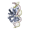
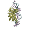
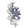
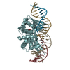
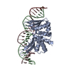
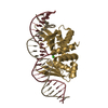
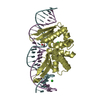
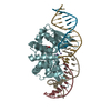
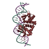
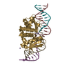
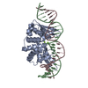
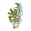
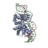
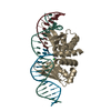
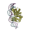
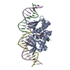
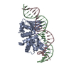
| タイトル | Structures and dynamics of mesophilic variants from the homing endonuclease I-DmoI | ||||||
 要素 要素 |
| ||||||
 キーワード キーワード | DNA BINDING PROTEIN / Desulfurococcus mobilis | ||||||
| 機能・相同性 |  機能・相同性情報 機能・相同性情報intron homing / intein-mediated protein splicing / endonuclease activity / 加水分解酵素; エステル加水分解酵素 / hydrolase activity 類似検索 - 分子機能 | ||||||
| 生物種 |  Desulfurococcus mucosus (古細菌) Desulfurococcus mucosus (古細菌)synthetic construct (人工物) | ||||||
| 手法 |  X線回折 / シンクロトロン / 分子置換 / 解像度: 2.75 Å X線回折 / シンクロトロン / 分子置換 / 解像度: 2.75 Å | ||||||
 データ登録者 データ登録者 | Molina, R. / Marcaida, M.J. | ||||||
 引用 引用 | ジャーナル: J. Comput. Aided Mol. Des. / 年: 2017 タイトル: Structure and dynamics of mesophilic variants from the homing endonuclease I-DmoI. 著者: Alba, J. / Marcaida, M.J. / Prieto, J. / Montoya, G. / Molina, R. / D'Abramo, M. | ||||||
| 履歴 |
|
- 構造の表示
構造の表示
| 構造ビューア | 分子: MolmilJmol/JSmol |
|---|
- ダウンロードとリンク
-ダウンロード
| PDBx/mmCIF形式 | 5o6g.cif.gz | 209.9 KB | 表示 | PDBx/mmCIF形式 |
|---|---|---|---|---|
| PDB形式 | pdb5o6g.ent.gz | 159.3 KB | 表示 | PDB形式 |
| PDBx/mmJSON形式 | 5o6g.json.gz | ツリー表示 | PDBx/mmJSON形式 | |
| その他 |  その他のダウンロード その他のダウンロード |
-検証レポート
| アーカイブディレクトリ | https://data.pdbj.org/pub/pdb/validation_reports/o6/5o6gftp://data.pdbj.org/pub/pdb/validation_reports/o6/5o6g | HTTPS FTP |
|---|
-関連構造データ

-リンク
 PDBj
PDBj


- 集合体
集合体
| 登録構造単位 | 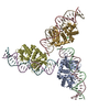
| ||||||||
|---|---|---|---|---|---|---|---|---|---|
| 1 | 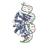
| ||||||||
| 2 | 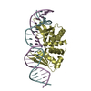
| ||||||||
| 3 | 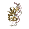
| ||||||||
| 単位格子 |
|
-要素
-タンパク質 , 1種, 3分子 ADG
| #1: タンパク質 | 分子量: 23445.240 Da / 分子数: 3 / 由来タイプ: 組換発現 / 由来: (組換発現) Desulfurococcus mucosus (古細菌) / 発現宿主:  参照: UniProt: P21505, 加水分解酵素; エステル加水分解酵素 |
|---|
-DNA鎖 , 2種, 6分子 BEHCFI
| #2: DNA鎖 | 分子量: 7707.932 Da / 分子数: 3 / 由来タイプ: 合成 / 由来: (合成) synthetic construct (人工物) #3: DNA鎖 | 分子量: 7654.926 Da / 分子数: 3 / 由来タイプ: 合成 / 由来: (合成) synthetic construct (人工物) |
|---|
-非ポリマー , 3種, 59分子 


| #4: 化合物 | ChemComp-MN /  分子量: 54.938 Da / 分子数: 6 / 由来タイプ: 組換発現 / 式: Mn 分子量: 54.938 Da / 分子数: 6 / 由来タイプ: 組換発現 / 式: Mn#5: 化合物 | ChemComp-CL / |  分子量: 35.453 Da / 分子数: 1 / 由来タイプ: 合成 / 式: Cl 分子量: 35.453 Da / 分子数: 1 / 由来タイプ: 合成 / 式: Cl#6: 水 | ChemComp-HOH / | 分子量: 18.015 Da / 分子数: 52 / 由来タイプ: 天然 / 式: H2O |
|---|
-実験情報
-実験
| 実験 | 手法: X線回折 / 使用した結晶の数: 1 |
|---|
- 試料調製
試料調製
| 結晶 | マシュー密度: 3.02 Å3/Da / 溶媒含有率: 59.24 % |
|---|---|
| 結晶化 | 温度: 293 K / 手法: 蒸気拡散法, シッティングドロップ法 詳細: 5-6 % PEG4000, 0.07 M NaAc pH = 4.6-5.5 and 30% Glycerol |
-データ収集
| 回折 | 平均測定温度: 100 K |
|---|---|
| 放射光源 | 由来: シンクロトロン / サイト: SLS  / ビームライン: X06SA / 波長: 1 Å / ビームライン: X06SA / 波長: 1 Å |
| 検出器 | タイプ: DECTRIS PILATUS3 6M / 検出器: PIXEL / 日付: 2010年6月6日 |
| 放射 | プロトコル: SINGLE WAVELENGTH / 単色(M)・ラウエ(L): M / 散乱光タイプ: x-ray |
| 放射波長 | 波長: 1 Å / 相対比: 1 |
| 反射 | 解像度: 2.75→93.1 Å / Num. obs: 35510 / % possible obs: 97.5 % / 冗長度: 4.7 % / Net I/σ(I): 9.4 |
- 解析
解析
| ソフトウェア |
| |||||||||||||||||||||||||||||||||||||||||||||||||||||||||||||||||||||||||||||||||||||||||||||||||||||||||
|---|---|---|---|---|---|---|---|---|---|---|---|---|---|---|---|---|---|---|---|---|---|---|---|---|---|---|---|---|---|---|---|---|---|---|---|---|---|---|---|---|---|---|---|---|---|---|---|---|---|---|---|---|---|---|---|---|---|---|---|---|---|---|---|---|---|---|---|---|---|---|---|---|---|---|---|---|---|---|---|---|---|---|---|---|---|---|---|---|---|---|---|---|---|---|---|---|---|---|---|---|---|---|---|---|---|---|
| 精密化 | 構造決定の手法: 分子置換 開始モデル: 4UN8 解像度: 2.75→56.184 Å / SU ML: 0.41 / 交差検証法: FREE R-VALUE / σ(F): 1.34 / 位相誤差: 24.82
| |||||||||||||||||||||||||||||||||||||||||||||||||||||||||||||||||||||||||||||||||||||||||||||||||||||||||
| 溶媒の処理 | 減衰半径: 0.9 Å / VDWプローブ半径: 1.11 Å | |||||||||||||||||||||||||||||||||||||||||||||||||||||||||||||||||||||||||||||||||||||||||||||||||||||||||
| 精密化ステップ | サイクル: LAST / 解像度: 2.75→56.184 Å
| |||||||||||||||||||||||||||||||||||||||||||||||||||||||||||||||||||||||||||||||||||||||||||||||||||||||||
| 拘束条件 |
| |||||||||||||||||||||||||||||||||||||||||||||||||||||||||||||||||||||||||||||||||||||||||||||||||||||||||
| LS精密化 シェル |
|
