 Movie
Movie Controller

[English] 日本語
 Yorodumi
- PDB-5o6i: Structures and dynamics of mesophilic variants from the homing en... -
+ Open data

- Basic information
| Entry | Database: PDB / ID: 5o6i | ||||||
|---|---|---|---|---|---|---|---|
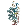
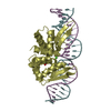
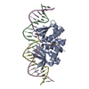
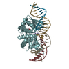
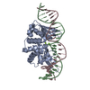
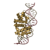
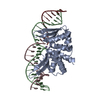
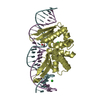
| Title | Structures and dynamics of mesophilic variants from the homing endonuclease I-DmoI | ||||||
 Components Components |
| ||||||
 Keywords Keywords | DNA BINDING PROTEIN / Desulfurococcus mobilis | ||||||
| Function / homology |  Function and homology information Function and homology informationintron homing / intein-mediated protein splicing / endonuclease activity / Hydrolases; Acting on ester bonds / hydrolase activity Similarity search - Function | ||||||
| Biological species |  Desulfurococcus mucosus (archaea) Desulfurococcus mucosus (archaea)synthetic construct (others) | ||||||
| Method |  X-RAY DIFFRACTION / SYNCHROTRON / MOLECULAR REPLACEMENT / Resolution: 2.25 Å X-RAY DIFFRACTION / SYNCHROTRON / MOLECULAR REPLACEMENT / Resolution: 2.25 Å | ||||||
 Authors Authors | Molina, R. / Marcaida, M.J. | ||||||
 Citation Citation | Journal: J. Comput. Aided Mol. Des. / Year: 2017 Title: Structure and dynamics of mesophilic variants from the homing endonuclease I-DmoI. Authors: Alba, J. / Marcaida, M.J. / Prieto, J. / Montoya, G. / Molina, R. / D'Abramo, M. | ||||||
| History |
|
- Structure visualization
| Structure viewer | Molecule: MolmilJmol/JSmol |
|---|
- Downloads & links
-Download
| PDBx/mmCIF format | 5o6i.cif.gz | 216.8 KB | Display | PDBx/mmCIF format |
|---|---|---|---|---|
| PDB format | pdb5o6i.ent.gz | 164.6 KB | Display | PDB format |
| PDBx/mmJSON format | 5o6i.json.gz | Tree view | PDBx/mmJSON format | |
| Others |  Other downloads Other downloads |
-Validation report
| Arichive directory | https://data.pdbj.org/pub/pdb/validation_reports/o6/5o6iftp://data.pdbj.org/pub/pdb/validation_reports/o6/5o6i | HTTPS FTP |
|---|
-Related structure data
| Related structure data |  5o6gC 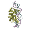 4un8S S: Starting model for refinement C: citing same article ( |
|---|---|
| Similar structure data |


-Links
 PDBj
PDBj


- Assembly
| Deposited unit | 
| ||||||||
|---|---|---|---|---|---|---|---|---|---|
| 1 | 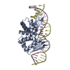
| ||||||||
| 2 | 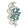
| ||||||||
| 3 | 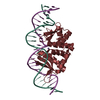
| ||||||||
| Unit cell |
|
-Components
-Protein , 1 types, 3 molecules AFK
| #1: Protein | Mass: 23416.266 Da / Num. of mol.: 3 Source method: isolated from a genetically manipulated source Source: (gene. exp.) Desulfurococcus mucosus (archaea) / Production host:  References: UniProt: P21505, Hydrolases; Acting on ester bonds |
|---|
-DNA chain , 2 types, 6 molecules CGLDIN
| #2: DNA chain | Mass: 7707.932 Da / Num. of mol.: 3 / Source method: obtained synthetically / Source: (synth.) synthetic construct (others) #3: DNA chain | Mass: 7654.926 Da / Num. of mol.: 3 / Source method: obtained synthetically / Source: (synth.) synthetic construct (others) |
|---|
-Non-polymers , 3 types, 291 molecules 


| #4: Chemical | ChemComp-MN /  Mass: 54.938 Da / Num. of mol.: 6 / Source method: obtained synthetically / Formula: Mn Mass: 54.938 Da / Num. of mol.: 6 / Source method: obtained synthetically / Formula: Mn#5: Chemical |  Mass: 35.453 Da / Num. of mol.: 2 / Source method: obtained synthetically / Formula: Cl Mass: 35.453 Da / Num. of mol.: 2 / Source method: obtained synthetically / Formula: Cl#6: Water | ChemComp-HOH / | Mass: 18.015 Da / Num. of mol.: 283 / Source method: isolated from a natural source / Formula: H2O |
|---|
-Experimental details
-Experiment
| Experiment | Method: X-RAY DIFFRACTION / Number of used crystals: 1 |
|---|
- Sample preparation
| Crystal | Density Matthews: 3.02 Å3/Da / Density % sol: 59.25 % |
|---|---|
| Crystal grow | Temperature: 293 K / Method: vapor diffusion, sitting drop Details: 5-6 % PEG4000, 0.07 M NaAc pH = 4.6-5.5 and 30% Glycerol |
-Data collection
| Diffraction | Mean temperature: 100 K |
|---|---|
| Diffraction source | Source: SYNCHROTRON / Site: SLS  / Beamline: X06SA / Wavelength: 1 Å / Beamline: X06SA / Wavelength: 1 Å |
| Detector | Type: DECTRIS PILATUS3 S 6M / Detector: PIXEL / Date: Jun 6, 2010 |
| Radiation | Protocol: SINGLE WAVELENGTH / Monochromatic (M) / Laue (L): M / Scattering type: x-ray |
| Radiation wavelength | Wavelength: 1 Å / Relative weight: 1 |
| Reflection | Resolution: 2.25→56.14 Å / Num. obs: 64736 / % possible obs: 98 % / Redundancy: 5.2 % / Net I/σ(I): 11.7 |
- Processing
| Software |
| ||||||||||||||||||||||||||||||||||||||||||||||||||||||||||||||||||||||||||||||||||||||||||||||||||||||||||||||||||||||||||||||||||||||||||||||||||||||||||||||||||||||||
|---|---|---|---|---|---|---|---|---|---|---|---|---|---|---|---|---|---|---|---|---|---|---|---|---|---|---|---|---|---|---|---|---|---|---|---|---|---|---|---|---|---|---|---|---|---|---|---|---|---|---|---|---|---|---|---|---|---|---|---|---|---|---|---|---|---|---|---|---|---|---|---|---|---|---|---|---|---|---|---|---|---|---|---|---|---|---|---|---|---|---|---|---|---|---|---|---|---|---|---|---|---|---|---|---|---|---|---|---|---|---|---|---|---|---|---|---|---|---|---|---|---|---|---|---|---|---|---|---|---|---|---|---|---|---|---|---|---|---|---|---|---|---|---|---|---|---|---|---|---|---|---|---|---|---|---|---|---|---|---|---|---|---|---|---|---|---|---|---|---|
| Refinement | Method to determine structure: MOLECULAR REPLACEMENT Starting model: 4UN8 Resolution: 2.25→56.137 Å / SU ML: 0.29 / Cross valid method: FREE R-VALUE / σ(F): 1.33 / Phase error: 25.42 / Stereochemistry target values: ML
| ||||||||||||||||||||||||||||||||||||||||||||||||||||||||||||||||||||||||||||||||||||||||||||||||||||||||||||||||||||||||||||||||||||||||||||||||||||||||||||||||||||||||
| Solvent computation | Shrinkage radii: 0.9 Å / VDW probe radii: 1.11 Å / Solvent model: FLAT BULK SOLVENT MODEL | ||||||||||||||||||||||||||||||||||||||||||||||||||||||||||||||||||||||||||||||||||||||||||||||||||||||||||||||||||||||||||||||||||||||||||||||||||||||||||||||||||||||||
| Refinement step | Cycle: LAST / Resolution: 2.25→56.137 Å
| ||||||||||||||||||||||||||||||||||||||||||||||||||||||||||||||||||||||||||||||||||||||||||||||||||||||||||||||||||||||||||||||||||||||||||||||||||||||||||||||||||||||||
| Refine LS restraints |
| ||||||||||||||||||||||||||||||||||||||||||||||||||||||||||||||||||||||||||||||||||||||||||||||||||||||||||||||||||||||||||||||||||||||||||||||||||||||||||||||||||||||||
| LS refinement shell |
|
