 Movie
Movie Controller

[English] 日本語
 Yorodumi
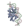
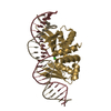
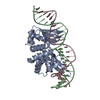
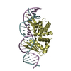
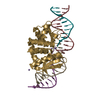
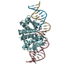
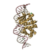
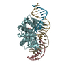
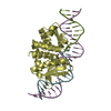
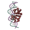
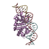
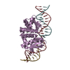
- PDB-4un9: THE CRYSTAL STRUCTURE OF I-DMOI IN COMPLEX WITH ITS TARGET DNA AT... -
+ Open data

- Basic information
| Entry | Database: PDB / ID: 4un9 | ||||||
|---|---|---|---|---|---|---|---|
| Title | THE CRYSTAL STRUCTURE OF I-DMOI IN COMPLEX WITH ITS TARGET DNA AT 8H INCUBATION IN 5MM MN (STATE 3) | ||||||
 Components Components |
| ||||||
 Keywords Keywords | HYDROLASE/DNA / HYDROLASE-DNA COMPLEX / CATALYSIS / PROTEIN-DNA INTERACTION | ||||||
| Function / homology |  Function and homology information Function and homology informationintron homing / intein-mediated protein splicing / endonuclease activity / Hydrolases; Acting on ester bonds / hydrolase activity Similarity search - Function | ||||||
| Biological species |  DESULFUROCOCCUS MOBILIS (archaea) DESULFUROCOCCUS MOBILIS (archaea)SYNTHETIC CONSTRUCT (others) | ||||||
| Method |  X-RAY DIFFRACTION / SYNCHROTRON / MOLECULAR REPLACEMENT / Resolution: 2.734 Å X-RAY DIFFRACTION / SYNCHROTRON / MOLECULAR REPLACEMENT / Resolution: 2.734 Å | ||||||
 Authors Authors | Molina, R. / Stella, S. / Redondo, P. / Gomez, H. / Marcaida, M.J. / Orozco, M. / Prieto, J. / Montoya, G. | ||||||
 Citation Citation | Journal: Nat.Struct.Mol.Biol. / Year: 2015 Title: Visualizing Phosphodiester-Bond Hydrolysis by an Endonuclease. Authors: Molina, R. / Stella, S. / Redondo, P. / Gomez, H. / Marcaida, M.J. / Orozco, M. / Prieto, J. / Montoya, G. | ||||||
| History |
|
- Structure visualization
| Structure viewer | Molecule: MolmilJmol/JSmol |
|---|
- Downloads & links
-Download
| PDBx/mmCIF format | 4un9.cif.gz | 211.9 KB | Display | PDBx/mmCIF format |
|---|---|---|---|---|
| PDB format | pdb4un9.ent.gz | 161.6 KB | Display | PDB format |
| PDBx/mmJSON format | 4un9.json.gz | Tree view | PDBx/mmJSON format | |
| Others |  Other downloads Other downloads |
-Validation report
| Arichive directory | https://data.pdbj.org/pub/pdb/validation_reports/un/4un9ftp://data.pdbj.org/pub/pdb/validation_reports/un/4un9 | HTTPS FTP |
|---|
-Related structure data
| Related structure data | 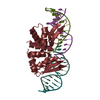 4d6nC 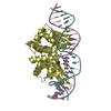 4d6oC  4un7C 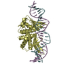 4un8C 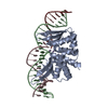 4unaC  4unbC 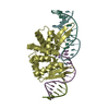 4uncC 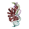 4ut0C 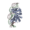 2vs7S  4un6 C: citing same article ( S: Starting model for refinement |
|---|---|
| Similar structure data |

-Links
 PDBj
PDBj


- Assembly
| Deposited unit | 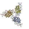
| ||||||||
|---|---|---|---|---|---|---|---|---|---|
| 1 | 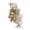
| ||||||||
| 2 | 
| ||||||||
| 3 | 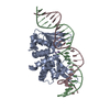
| ||||||||
| Unit cell |
|
-Components
-HOMING ENDONUCLEASE I- ... , 2 types, 3 molecules ADG
| #1: Protein | Mass: 23265.057 Da / Num. of mol.: 2 Source method: isolated from a genetically manipulated source Source: (gene. exp.) DESULFUROCOCCUS MOBILIS (archaea) / Production host:  References: UniProt: P21505, Hydrolases; Acting on ester bonds #4: Protein | | Mass: 23280.068 Da / Num. of mol.: 1 Source method: isolated from a genetically manipulated source Source: (gene. exp.) DESULFUROCOCCUS MOBILIS (archaea) / Production host: References: UniProt: P21505, Hydrolases; Acting on ester bonds |
|---|
-DNA chain , 2 types, 6 molecules BEHCFI
| #2: DNA chain | Mass: 7707.932 Da / Num. of mol.: 3 / Source method: obtained synthetically / Source: (synth.) SYNTHETIC CONSTRUCT (others) #3: DNA chain | Mass: 7654.926 Da / Num. of mol.: 3 / Source method: obtained synthetically / Source: (synth.) SYNTHETIC CONSTRUCT (others) |
|---|
-Non-polymers , 2 types, 114 molecules 

| #5: Chemical | ChemComp-MN /  Mass: 54.938 Da / Num. of mol.: 10 / Source method: obtained synthetically / Formula: Mn Mass: 54.938 Da / Num. of mol.: 10 / Source method: obtained synthetically / Formula: Mn#6: Water | ChemComp-HOH / | Mass: 18.015 Da / Num. of mol.: 104 / Source method: isolated from a natural source / Formula: H2O |
|---|
-Experimental details
-Experiment
| Experiment | Method: X-RAY DIFFRACTION / Number of used crystals: 1 |
|---|
- Sample preparation
| Crystal | Density Matthews: 2.31 Å3/Da / Density % sol: 52 % / Description: NONE |
|---|---|
| Crystal grow | pH: 6 / Details: pH 6 |
-Data collection
| Diffraction | Mean temperature: 100 K |
|---|---|
| Diffraction source | Source: SYNCHROTRON / Site: SLS  / Beamline: X06SA / Wavelength: 1.8 / Beamline: X06SA / Wavelength: 1.8 |
| Detector | Type: DECTRIS PILATUS 6M / Detector: PIXEL |
| Radiation | Protocol: SINGLE WAVELENGTH / Monochromatic (M) / Laue (L): M / Scattering type: x-ray |
| Radiation wavelength | Wavelength: 1.8 Å / Relative weight: 1 |
| Reflection | Resolution: 2.73→46.65 Å / Num. obs: 39637 / % possible obs: 99.9 % / Observed criterion σ(I): 2 / Redundancy: 4.7 % / Biso Wilson estimate: 55.12 Å2 / Rmerge(I) obs: 0.11 / Net I/σ(I): 10.5 |
| Reflection shell | Resolution: 2.73→2.85 Å / Redundancy: 4.6 % / Rmerge(I) obs: 0.57 / Mean I/σ(I) obs: 2.3 / % possible all: 99.9 |
- Processing
| Software |
| ||||||||||||||||||||||||||||||||||||||||||||||||||||||||||||||||||||||||||||||||||||||||||||||||||||||||||||||||||||||||||||||||||||||||||||||||||||||||||||||||||||||||||||||||||||||
|---|---|---|---|---|---|---|---|---|---|---|---|---|---|---|---|---|---|---|---|---|---|---|---|---|---|---|---|---|---|---|---|---|---|---|---|---|---|---|---|---|---|---|---|---|---|---|---|---|---|---|---|---|---|---|---|---|---|---|---|---|---|---|---|---|---|---|---|---|---|---|---|---|---|---|---|---|---|---|---|---|---|---|---|---|---|---|---|---|---|---|---|---|---|---|---|---|---|---|---|---|---|---|---|---|---|---|---|---|---|---|---|---|---|---|---|---|---|---|---|---|---|---|---|---|---|---|---|---|---|---|---|---|---|---|---|---|---|---|---|---|---|---|---|---|---|---|---|---|---|---|---|---|---|---|---|---|---|---|---|---|---|---|---|---|---|---|---|---|---|---|---|---|---|---|---|---|---|---|---|---|---|---|---|
| Refinement | Method to determine structure: MOLECULAR REPLACEMENT Starting model: PDB ENTRY 2VS7 Resolution: 2.734→42.86 Å / SU ML: 0.38 / σ(F): 1.2 / Phase error: 22.99 / Stereochemistry target values: ML
| ||||||||||||||||||||||||||||||||||||||||||||||||||||||||||||||||||||||||||||||||||||||||||||||||||||||||||||||||||||||||||||||||||||||||||||||||||||||||||||||||||||||||||||||||||||||
| Solvent computation | Shrinkage radii: 0.9 Å / VDW probe radii: 1.11 Å / Solvent model: FLAT BULK SOLVENT MODEL | ||||||||||||||||||||||||||||||||||||||||||||||||||||||||||||||||||||||||||||||||||||||||||||||||||||||||||||||||||||||||||||||||||||||||||||||||||||||||||||||||||||||||||||||||||||||
| Refinement step | Cycle: LAST / Resolution: 2.734→42.86 Å
| ||||||||||||||||||||||||||||||||||||||||||||||||||||||||||||||||||||||||||||||||||||||||||||||||||||||||||||||||||||||||||||||||||||||||||||||||||||||||||||||||||||||||||||||||||||||
| Refine LS restraints |
| ||||||||||||||||||||||||||||||||||||||||||||||||||||||||||||||||||||||||||||||||||||||||||||||||||||||||||||||||||||||||||||||||||||||||||||||||||||||||||||||||||||||||||||||||||||||
| LS refinement shell |
|
