 Movie
Movie Controller


+ Open data

- Basic information
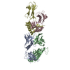
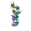
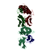
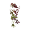
| Entry | Database: PDB / ID: 7bxo | ||||||
|---|---|---|---|---|---|---|---|
| Title | Crystal structure of the toxin-antitoxin with AMP-PNP | ||||||
 Components Components |
| ||||||
 Keywords Keywords | TOXIN / a novel Toxin-Antitoxin system | ||||||
| Function / homology |  Function and homology information Function and homology informationAMPylase activity / protein adenylyltransferase / toxin-antitoxin complex / RNA nuclease activity / endonuclease activity / Hydrolases; Acting on ester bonds / nucleotide binding / hydrolase activity / DNA binding / ATP binding / metal ion binding Similarity search - Function | ||||||
| Biological species |  Shewanella oneidensis (bacteria) Shewanella oneidensis (bacteria) | ||||||
| Method |  X-RAY DIFFRACTION / SYNCHROTRON / MOLECULAR REPLACEMENT / Resolution: 2.77 Å X-RAY DIFFRACTION / SYNCHROTRON / MOLECULAR REPLACEMENT / Resolution: 2.77 Å | ||||||
 Authors Authors | Ouyang, S.Y. / Zhen, X.K. | ||||||
 Citation Citation | Journal: Nucleic Acids Res. / Year: 2020 Title: Novel polyadenylylation-dependent neutralization mechanism of the HEPN/MNT toxin/antitoxin system. Authors: Yao, J. / Zhen, X. / Tang, K. / Liu, T. / Xu, X. / Chen, Z. / Guo, Y. / Liu, X. / Wood, T.K. / Ouyang, S. / Wang, X. | ||||||
| History |
|
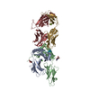
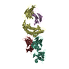
- Structure visualization
| Structure viewer | Molecule: MolmilJmol/JSmol |
|---|
- Downloads & links
-Download
| PDBx/mmCIF format | 7bxo.cif.gz | 214.3 KB | Display | PDBx/mmCIF format |
|---|---|---|---|---|
| PDB format | pdb7bxo.ent.gz | 170.9 KB | Display | PDB format |
| PDBx/mmJSON format | 7bxo.json.gz | Tree view | PDBx/mmJSON format | |
| Others |  Other downloads Other downloads |
-Validation report
| Arichive directory | https://data.pdbj.org/pub/pdb/validation_reports/bx/7bxoftp://data.pdbj.org/pub/pdb/validation_reports/bx/7bxo | HTTPS FTP |
|---|
-Related structure data
| Related structure data |  6m6uC  6m6vC  6m6wC  5yepS S: Starting model for refinement C: citing same article ( |
|---|---|
| Similar structure data |


-Links
 PDBj
PDBj


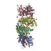
- Assembly
| Deposited unit | 
| ||||||||
|---|---|---|---|---|---|---|---|---|---|
| 1 |
| ||||||||
| Unit cell |
|
-Components
| #1: Protein | Mass: 15582.584 Da / Num. of mol.: 2 Source method: isolated from a genetically manipulated source Source: (gene. exp.) Shewanella oneidensis (strain MR-1) (bacteria)Strain: MR-1 / Gene: SO_3165 / Production host: #2: Protein | Mass: 15238.448 Da / Num. of mol.: 6 / Mutation: Y104A Source method: isolated from a genetically manipulated source Source: (gene. exp.) Shewanella oneidensis (strain MR-1) (bacteria)Strain: MR-1 / Gene: SO_3166 / Production host: #3: Chemical |   Mass: 506.196 Da / Num. of mol.: 2 / Source method: obtained synthetically / Formula: C10H17N6O12P3 / Feature type: SUBJECT OF INVESTIGATION / Comment: AMP-PNP, energy-carrying molecule analogue*YM Mass: 506.196 Da / Num. of mol.: 2 / Source method: obtained synthetically / Formula: C10H17N6O12P3 / Feature type: SUBJECT OF INVESTIGATION / Comment: AMP-PNP, energy-carrying molecule analogue*YM#4: Chemical | ChemComp-MG /   Mass: 24.305 Da / Num. of mol.: 4 / Source method: obtained synthetically / Formula: Mg / Feature type: SUBJECT OF INVESTIGATION Mass: 24.305 Da / Num. of mol.: 4 / Source method: obtained synthetically / Formula: Mg / Feature type: SUBJECT OF INVESTIGATION#5: Water | ChemComp-HOH / |  Mass: 18.015 Da / Num. of mol.: 14 / Source method: isolated from a natural source / Formula: H2O Mass: 18.015 Da / Num. of mol.: 14 / Source method: isolated from a natural source / Formula: H2OHas ligand of interest | Y | |
|---|
-Experimental details
-Experiment
| Experiment | Method: X-RAY DIFFRACTION / Number of used crystals: 1 |
|---|
- Sample preparation
| Crystal | Density Matthews: 2.93 Å3/Da / Density % sol: 58.08 % |
|---|---|
| Crystal grow | Temperature: 291 K / Method: vapor diffusion, sitting drop / pH: 8 Details: 0.1 M Tris-HCl , 0.2 M potassium sodium tartrate, and 12-20% (v/v) PEG 3350 PH range: pH 7.8-8.2 |
-Data collection
| Diffraction | Mean temperature: 100 K / Serial crystal experiment: N |
|---|---|
| Diffraction source | Source: SYNCHROTRON / Site: SSRF  / Beamline: BL17U1 / Wavelength: 0.9792 Å / Beamline: BL17U1 / Wavelength: 0.9792 Å |
| Detector | Type: DECTRIS EIGER X 16M / Detector: PIXEL / Date: Oct 10, 2019 |
| Radiation | Protocol: SINGLE WAVELENGTH / Monochromatic (M) / Laue (L): M / Scattering type: x-ray |
| Radiation wavelength | Wavelength: 0.9792 Å / Relative weight: 1 |
| Reflection | Resolution: 2.77→100.68 Å / Num. obs: 36307 / % possible obs: 99.9 % / Redundancy: 6.8 % / Biso Wilson estimate: 94.42 Å2 / CC1/2: 0.999 / Net I/σ(I): 15.9 |
| Reflection shell | Resolution: 2.77→2.86 Å / Num. unique obs: 2668 / CC1/2: 0.999 / % possible all: 99.9 |
- Processing
| Software |
| ||||||||||||||||||||||||
|---|---|---|---|---|---|---|---|---|---|---|---|---|---|---|---|---|---|---|---|---|---|---|---|---|---|
| Refinement | Method to determine structure: MOLECULAR REPLACEMENT Starting model: 5YEP Resolution: 2.77→36.14 Å / Cor.coef. Fo:Fc: 0.939 / Cor.coef. Fo:Fc free: 0.933 / SU R Cruickshank DPI: 1.035 / Cross valid method: THROUGHOUT / σ(F): 0 / SU R Blow DPI: 0.8 / SU Rfree Blow DPI: 0.318 / SU Rfree Cruickshank DPI: 0.331
| ||||||||||||||||||||||||
| Displacement parameters | Biso max: 172.96 Å2 / Biso mean: 86.49 Å2 / Biso min: 38.12 Å2
| ||||||||||||||||||||||||
| Refine analyze | Luzzati coordinate error obs: 0.41 Å | ||||||||||||||||||||||||
| Refinement step | Cycle: final / Resolution: 2.77→36.14 Å
| ||||||||||||||||||||||||
| LS refinement shell | Resolution: 2.77→2.79 Å / Rfactor Rfree error: 0 / Total num. of bins used: 50
|
