 Movie
Movie Controller

[English] 日本語
 Yorodumi
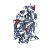
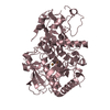
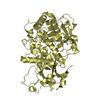
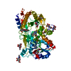
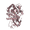
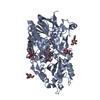
- PDB-7bwp: Crystal complex of endo-deglycosylated PcHNL5 with (R)-mandelonitrile -
+ Open data

- Basic information
| Entry | Database: PDB / ID: 7bwp | ||||||
|---|---|---|---|---|---|---|---|
| Title | Crystal complex of endo-deglycosylated PcHNL5 with (R)-mandelonitrile | ||||||
 Components Components | PREDICTED: (R)-mandelonitrile lyase | ||||||
 Keywords Keywords | LYASE / Complex / endo-deglycosylated | ||||||
| Function / homology |  Function and homology information Function and homology information(R)-mandelonitrile lyase / mandelonitrile lyase activity / oxidoreductase activity, acting on CH-OH group of donors / flavin adenine dinucleotide binding Similarity search - Function | ||||||
| Biological species |  | ||||||
| Method |  X-RAY DIFFRACTION / SYNCHROTRON / MOLECULAR REPLACEMENT / Resolution: 1.802 Å X-RAY DIFFRACTION / SYNCHROTRON / MOLECULAR REPLACEMENT / Resolution: 1.802 Å | ||||||
 Authors Authors | Zheng, Y.C. / Li, F.L. / Yu, H.L. / Xu, J.H. | ||||||
 Citation Citation | Journal: Acs Catalysis / Year: 2020 Title: Structure-Guided Tuning of a Hydroxynitrile Lyase to Accept Rigid Pharmaco Aldehydes. Authors: Zheng, Y.C. / Li, F.L. / Lin, Z.M. / Lin, G.Q. / Hong, R. / Yu, H.L. / Xu, J.H. | ||||||
| History |
|
- Structure visualization
| Structure viewer | Molecule: MolmilJmol/JSmol |
|---|
- Downloads & links
-Download
| PDBx/mmCIF format | 7bwp.cif.gz | 132 KB | Display | PDBx/mmCIF format |
|---|---|---|---|---|
| PDB format | pdb7bwp.ent.gz | 96.2 KB | Display | PDB format |
| PDBx/mmJSON format | 7bwp.json.gz | Tree view | PDBx/mmJSON format | |
| Others |  Other downloads Other downloads |
-Validation report
| Arichive directory | https://data.pdbj.org/pub/pdb/validation_reports/bw/7bwpftp://data.pdbj.org/pub/pdb/validation_reports/bw/7bwp | HTTPS FTP |
|---|
-Related structure data
| Related structure data |  6jbySC 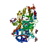 6lqyC  6lr8C S: Starting model for refinement C: citing same article ( |
|---|---|
| Similar structure data |


-Links
 PDBj
PDBj

- Assembly
| Deposited unit | 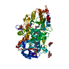
| ||||||||
|---|---|---|---|---|---|---|---|---|---|
| 1 |
| ||||||||
| Unit cell |
|
-Components
-Protein / Sugars , 2 types, 7 molecules A
| #1: Protein | Mass: 59186.566 Da / Num. of mol.: 1 Source method: isolated from a genetically manipulated source Source: (gene. exp.)  Komagataella pastoris (fungus) / References: UniProt: A0A5E4GBK6, UniProt: O24243*PLUS Komagataella pastoris (fungus) / References: UniProt: A0A5E4GBK6, UniProt: O24243*PLUS |
|---|---|
| #3: Sugar | ChemComp-NAG /  Type: D-saccharide, beta linking / Mass: 221.208 Da / Num. of mol.: 6 Type: D-saccharide, beta linking / Mass: 221.208 Da / Num. of mol.: 6Source method: isolated from a genetically manipulated source Formula: C8H15NO6 |
-Non-polymers , 5 types, 452 molecules 


| #2: Chemical | ChemComp-MXN / ( Mass: 133.147 Da / Num. of mol.: 1 / Source method: obtained synthetically / Formula: C8H7NO / Feature type: SUBJECT OF INVESTIGATION Mass: 133.147 Da / Num. of mol.: 1 / Source method: obtained synthetically / Formula: C8H7NO / Feature type: SUBJECT OF INVESTIGATION |
|---|---|
| #4: Chemical | ChemComp-FAD /  Mass: 785.550 Da / Num. of mol.: 1 / Source method: obtained synthetically / Formula: C27H33N9O15P2 / Comment: FAD*YM Mass: 785.550 Da / Num. of mol.: 1 / Source method: obtained synthetically / Formula: C27H33N9O15P2 / Comment: FAD*YM |
| #5: Chemical | ChemComp-PO4 /  Mass: 94.971 Da / Num. of mol.: 1 / Source method: obtained synthetically / Formula: PO4 Mass: 94.971 Da / Num. of mol.: 1 / Source method: obtained synthetically / Formula: PO4 |
| #6: Chemical | ChemComp-PEG /  Mass: 106.120 Da / Num. of mol.: 1 / Source method: obtained synthetically / Formula: C4H10O3 Mass: 106.120 Da / Num. of mol.: 1 / Source method: obtained synthetically / Formula: C4H10O3 |
| #7: Water | ChemComp-HOH / Mass: 18.015 Da / Num. of mol.: 448 / Source method: isolated from a natural source / Formula: H2O |
-Details
| Has ligand of interest | Y |
|---|---|
| Has protein modification | Y |
-Experimental details
-Experiment
| Experiment | Method: X-RAY DIFFRACTION / Number of used crystals: 1 |
|---|
- Sample preparation
| Crystal | Density Matthews: 2.48 Å3/Da / Density % sol: 50.44 % |
|---|---|
| Crystal grow | Temperature: 289 K / Method: vapor diffusion, sitting drop / pH: 8.25 Details: tris-bicine, 100 mM, pH 8.25; CaCl2, 60 mM; MgCl2, 60 mM; PEG 500MME, 24%, v/v; PEG 20000, 12%, w/v PH range: 8.25 - 8.5 |
-Data collection
| Diffraction | Mean temperature: 100 K / Serial crystal experiment: N |
|---|---|
| Diffraction source | Source: SYNCHROTRON / Site: SSRF  / Beamline: BL19U1 / Wavelength: 0.9789 Å / Beamline: BL19U1 / Wavelength: 0.9789 Å |
| Detector | Type: DECTRIS PILATUS3 6M / Detector: PIXEL / Date: Jun 22, 2018 |
| Radiation | Protocol: SINGLE WAVELENGTH / Monochromatic (M) / Laue (L): M / Scattering type: x-ray |
| Radiation wavelength | Wavelength: 0.9789 Å / Relative weight: 1 |
| Reflection | Resolution: 1.8→50 Å / Num. obs: 55236 / % possible obs: 99.8 % / Redundancy: 12.6 % / Biso Wilson estimate: 17.76 Å2 / CC1/2: 0.996 / Rrim(I) all: 0.057 / Net I/σ(I): 34.3 |
| Reflection shell | Resolution: 1.8→1.83 Å / Mean I/σ(I) obs: 16.6 / Num. unique obs: 2751 / CC1/2: 0.978 |
- Processing
| Software |
| ||||||||||||||||||||||||||||||||||||||||||||||||||||||||||||||||||||||||||||||||||||||||||||||||||||||||||||||||||||||||||||||
|---|---|---|---|---|---|---|---|---|---|---|---|---|---|---|---|---|---|---|---|---|---|---|---|---|---|---|---|---|---|---|---|---|---|---|---|---|---|---|---|---|---|---|---|---|---|---|---|---|---|---|---|---|---|---|---|---|---|---|---|---|---|---|---|---|---|---|---|---|---|---|---|---|---|---|---|---|---|---|---|---|---|---|---|---|---|---|---|---|---|---|---|---|---|---|---|---|---|---|---|---|---|---|---|---|---|---|---|---|---|---|---|---|---|---|---|---|---|---|---|---|---|---|---|---|---|---|---|
| Refinement | Method to determine structure: MOLECULAR REPLACEMENT Starting model: 6JBY Resolution: 1.802→46.305 Å / SU ML: 0.16 / Cross valid method: FREE R-VALUE / σ(F): 1.38 / Phase error: 16.98
| ||||||||||||||||||||||||||||||||||||||||||||||||||||||||||||||||||||||||||||||||||||||||||||||||||||||||||||||||||||||||||||||
| Solvent computation | Shrinkage radii: 0.9 Å / VDW probe radii: 1.11 Å | ||||||||||||||||||||||||||||||||||||||||||||||||||||||||||||||||||||||||||||||||||||||||||||||||||||||||||||||||||||||||||||||
| Displacement parameters | Biso max: 63.49 Å2 / Biso mean: 19.2832 Å2 / Biso min: 7.31 Å2 | ||||||||||||||||||||||||||||||||||||||||||||||||||||||||||||||||||||||||||||||||||||||||||||||||||||||||||||||||||||||||||||||
| Refinement step | Cycle: final / Resolution: 1.802→46.305 Å
| ||||||||||||||||||||||||||||||||||||||||||||||||||||||||||||||||||||||||||||||||||||||||||||||||||||||||||||||||||||||||||||||
| Refine LS restraints |
| ||||||||||||||||||||||||||||||||||||||||||||||||||||||||||||||||||||||||||||||||||||||||||||||||||||||||||||||||||||||||||||||
| LS refinement shell | Refine-ID: X-RAY DIFFRACTION / Rfactor Rfree error: 0
|
