 Movie
Movie Controller

[English] 日本語
 Yorodumi
- PDB-5eb4: The crystal structure of almond HNL, PaHNL5 V317A, expressed in A... -
+ Open data

- Basic information
| Entry | Database: PDB / ID: 5eb4 | |||||||||
|---|---|---|---|---|---|---|---|---|---|---|
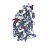
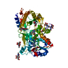
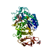
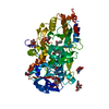
| Title | The crystal structure of almond HNL, PaHNL5 V317A, expressed in Aspergillus niger | |||||||||
 Components Components | Hnl isoenzyme 5 | |||||||||
 Keywords Keywords | LYASE / hydroxynitrile lyase / Prunus amygdalus / Aspergillus niger | |||||||||
| Function / homology |  Function and homology information Function and homology information(R)-mandelonitrile lyase / mandelonitrile lyase activity / oxidoreductase activity, acting on CH-OH group of donors / flavin adenine dinucleotide binding Similarity search - Function | |||||||||
| Biological species |  | |||||||||
| Method |  X-RAY DIFFRACTION / SYNCHROTRON / MOLECULAR REPLACEMENT / Resolution: 2.3 Å X-RAY DIFFRACTION / SYNCHROTRON / MOLECULAR REPLACEMENT / Resolution: 2.3 Å | |||||||||
 Authors Authors | Pavkov-Keller, T. / Steinkellner, G. / Gruber, K. | |||||||||
| Funding support |  Austria, 1items Austria, 1items
| |||||||||
 Citation Citation | Journal: J.Biotechnol. / Year: 2016 Title: Structures of almond hydroxynitrile lyase isoenzyme 5 provide a rationale for the lack of oxidoreductase activity in flavin dependent HNLs. Authors: Pavkov-Keller, T. / Bakhuis, J. / Steinkellner, G. / Jolink, F. / Keijmel, E. / Birner-Gruenberger, R. / Gruber, K. | |||||||||
| History |
|
- Structure visualization
| Structure viewer | Molecule: MolmilJmol/JSmol |
|---|
- Downloads & links
-Download
| PDBx/mmCIF format | 5eb4.cif.gz | 226.4 KB | Display | PDBx/mmCIF format |
|---|---|---|---|---|
| PDB format | pdb5eb4.ent.gz | 178.8 KB | Display | PDB format |
| PDBx/mmJSON format | 5eb4.json.gz | Tree view | PDBx/mmJSON format | |
| Others |  Other downloads Other downloads |
-Validation report
| Arichive directory | https://data.pdbj.org/pub/pdb/validation_reports/eb/5eb4ftp://data.pdbj.org/pub/pdb/validation_reports/eb/5eb4 | HTTPS FTP |
|---|
-Related structure data
| Related structure data |  5eb5C  1ju2S S: Starting model for refinement C: citing same article ( |
|---|---|
| Similar structure data |


-Links
 PDBj
PDBj

- Assembly
| Deposited unit | 
| ||||||||
|---|---|---|---|---|---|---|---|---|---|
| 1 | 
| ||||||||
| 2 | 
| ||||||||
| Unit cell |
|
-Components
| #1: Protein | Mass: 57903.172 Da / Num. of mol.: 2 / Fragment: UNP residues 28-559 / Mutation: V317A Source method: isolated from a genetically manipulated source Source: (gene. exp.)  #2: Polysaccharide | Source method: isolated from a genetically manipulated source #3: Sugar | ChemComp-NAG /   Type: D-saccharide, beta linking / Mass: 221.208 Da / Num. of mol.: 10 Type: D-saccharide, beta linking / Mass: 221.208 Da / Num. of mol.: 10Source method: isolated from a genetically manipulated source Formula: C8H15NO6 #4: Chemical |   Mass: 785.550 Da / Num. of mol.: 2 / Source method: obtained synthetically / Formula: C27H33N9O15P2 / Comment: FAD*YM Mass: 785.550 Da / Num. of mol.: 2 / Source method: obtained synthetically / Formula: C27H33N9O15P2 / Comment: FAD*YM#5: Water | ChemComp-HOH / |  Mass: 18.015 Da / Num. of mol.: 464 / Source method: isolated from a natural source / Formula: H2O Mass: 18.015 Da / Num. of mol.: 464 / Source method: isolated from a natural source / Formula: H2OHas protein modification | Y | |
|---|
-Experimental details
-Experiment
| Experiment | Method: X-RAY DIFFRACTION |
|---|
- Sample preparation
| Crystal | Density Matthews: 2.67 Å3/Da / Density % sol: 53.99 % |
|---|---|
| Crystal grow | Temperature: 298 K / Method: vapor diffusion, sitting drop Details: The initial protein concentration was 28 mg/ml (10mM L-Malic acid, MES, Tris pH 7). Different crystal forms were obtained under crystallization conditions varying - PEG 4K, isopropanol and ...Details: The initial protein concentration was 28 mg/ml (10mM L-Malic acid, MES, Tris pH 7). Different crystal forms were obtained under crystallization conditions varying - PEG 4K, isopropanol and 10mM Hepes pH 7-7.8 with protein concentration 19-28 mg/ml. Most of the crystals that appeared within 2 weeks had visual grow defects. In one of the drops the stack of thin plates appeared after 1 month and one complete dataset was collected. PH range: 7.0 - 7.8 |
-Data collection
| Diffraction | Mean temperature: 100 K Ambient temp details: data were collected at the microfocus beamline (SLS) in 3 passes, since the automatic centering of the small thin, plate-like crystal was difficult |
|---|---|
| Diffraction source | Source: SYNCHROTRON / Site: SLS  / Beamline: X06SA / Wavelength: 1 Å / Beamline: X06SA / Wavelength: 1 Å |
| Detector | Type: MARMOSAIC 225 mm CCD / Detector: CCD / Date: May 29, 2011 |
| Radiation | Protocol: SINGLE WAVELENGTH / Monochromatic (M) / Laue (L): M / Scattering type: x-ray |
| Radiation wavelength | Wavelength: 1 Å / Relative weight: 1 |
| Reflection | Resolution: 2.3→49.1 Å / Num. obs: 49502 / % possible obs: 91.4 % / Redundancy: 5.3 % / Rmerge(I) obs: 0.188 / Net I/σ(I): 6.6 |
| Reflection shell | Resolution: 2.3→2.35 Å / Redundancy: 3.6 % / Rmerge(I) obs: 0.566 / Mean I/σ(I) obs: 2.5 / % possible all: 85.2 |
- Processing
| Software |
| ||||||||||||||||||||||||||||||||||||||||||||||||||||||||||||||||||||||||||||||||||||||||||||||||||||||||||||||||||||||||||||||||||||||||||||||||||||||||||||||||||||||||||||||||||||||
|---|---|---|---|---|---|---|---|---|---|---|---|---|---|---|---|---|---|---|---|---|---|---|---|---|---|---|---|---|---|---|---|---|---|---|---|---|---|---|---|---|---|---|---|---|---|---|---|---|---|---|---|---|---|---|---|---|---|---|---|---|---|---|---|---|---|---|---|---|---|---|---|---|---|---|---|---|---|---|---|---|---|---|---|---|---|---|---|---|---|---|---|---|---|---|---|---|---|---|---|---|---|---|---|---|---|---|---|---|---|---|---|---|---|---|---|---|---|---|---|---|---|---|---|---|---|---|---|---|---|---|---|---|---|---|---|---|---|---|---|---|---|---|---|---|---|---|---|---|---|---|---|---|---|---|---|---|---|---|---|---|---|---|---|---|---|---|---|---|---|---|---|---|---|---|---|---|---|---|---|---|---|---|---|
| Refinement | Method to determine structure: MOLECULAR REPLACEMENT Starting model: 1ju2 Resolution: 2.3→49.09 Å / Cor.coef. Fo:Fc: 0.958 / Cor.coef. Fo:Fc free: 0.917 / SU B: 9.043 / SU ML: 0.212 / Cross valid method: THROUGHOUT / ESU R: 0.373 / ESU R Free: 0.255 / Stereochemistry target values: MAXIMUM LIKELIHOOD / Details: HYDROGENS HAVE BEEN USED IF PRESENT IN THE INPUT
| ||||||||||||||||||||||||||||||||||||||||||||||||||||||||||||||||||||||||||||||||||||||||||||||||||||||||||||||||||||||||||||||||||||||||||||||||||||||||||||||||||||||||||||||||||||||
| Solvent computation | Ion probe radii: 0.8 Å / Shrinkage radii: 0.8 Å / VDW probe radii: 1.2 Å / Solvent model: MASK | ||||||||||||||||||||||||||||||||||||||||||||||||||||||||||||||||||||||||||||||||||||||||||||||||||||||||||||||||||||||||||||||||||||||||||||||||||||||||||||||||||||||||||||||||||||||
| Displacement parameters | Biso mean: 25.658 Å2
| ||||||||||||||||||||||||||||||||||||||||||||||||||||||||||||||||||||||||||||||||||||||||||||||||||||||||||||||||||||||||||||||||||||||||||||||||||||||||||||||||||||||||||||||||||||||
| Refinement step | Cycle: 1 / Resolution: 2.3→49.09 Å
| ||||||||||||||||||||||||||||||||||||||||||||||||||||||||||||||||||||||||||||||||||||||||||||||||||||||||||||||||||||||||||||||||||||||||||||||||||||||||||||||||||||||||||||||||||||||
| Refine LS restraints |
|