 Movie
Movie Controller

[English] 日本語
 Yorodumi
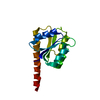
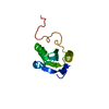
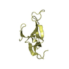
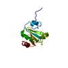
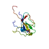
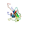
- PDB-7bmv: p62PH in cesium chloride (0.25 M CsCl in protein buffer and cryo ... -
+ Open data

- Basic information
| Entry | Database: PDB / ID: 7bmv | ||||||
|---|---|---|---|---|---|---|---|
| Title | p62PH in cesium chloride (0.25 M CsCl in protein buffer and cryo protectant) | ||||||
 Components Components | RNA polymerase II transcription factor B 73 kDa subunit-like protein | ||||||
 Keywords Keywords | TRANSCRIPTION / Cesium / Phasing / Pleckstrin homology domain | ||||||
| Function / homology |  Function and homology information Function and homology informationtranscription factor TFIIH core complex / nucleotide-excision repair / DNA-templated transcription Similarity search - Function | ||||||
| Biological species |  Chaetomium thermophilum (fungus) Chaetomium thermophilum (fungus) | ||||||
| Method |  X-RAY DIFFRACTION / SYNCHROTRON / SAD / Resolution: 1.9 Å X-RAY DIFFRACTION / SYNCHROTRON / SAD / Resolution: 1.9 Å | ||||||
 Authors Authors | Koelmel, W. / Kuper, J. / Kisker, C. | ||||||
| Funding support |  Germany, 1items Germany, 1items
| ||||||
 Citation Citation | Journal: Sci Rep / Year: 2021 Title: Cesium based phasing of macromolecules: a general easy to use approach for solving the phase problem. Authors: Koelmel, W. / Kuper, J. / Kisker, C. | ||||||
| History |
|
- Structure visualization
| Structure viewer | Molecule: MolmilJmol/JSmol |
|---|
- Downloads & links
-Download
| PDBx/mmCIF format | 7bmv.cif.gz | 38.6 KB | Display | PDBx/mmCIF format |
|---|---|---|---|---|
| PDB format | pdb7bmv.ent.gz | 24.7 KB | Display | PDB format |
| PDBx/mmJSON format | 7bmv.json.gz | Tree view | PDBx/mmJSON format | |
| Others |  Other downloads Other downloads |
-Validation report
| Summary document | 7bmv_validation.pdf.gz | 980.7 KB | Display | wwPDB validaton report |
|---|---|---|---|---|
| Full document | 7bmv_full_validation.pdf.gz | 980.7 KB | Display | |
| Data in XML | 7bmv_validation.xml.gz | 7.2 KB | Display | |
| Data in CIF | 7bmv_validation.cif.gz | 9.6 KB | Display | |
| Arichive directory | https://data.pdbj.org/pub/pdb/validation_reports/bm/7bmvftp://data.pdbj.org/pub/pdb/validation_reports/bm/7bmv | HTTPS FTP |
-Related structure data
| Related structure data | 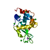 7bmoC  7bmpC 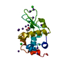 7bmqC 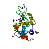 7bmrC 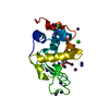 7bmsC 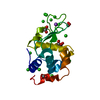 7bmtC 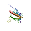 7bmuC 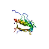 7bmwC 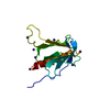 7bmxC 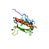 7bmyC 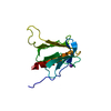 7bmzC C: citing same article ( |
|---|---|
| Similar structure data |


-Links
 PDBj
PDBj- Assembly
| Deposited unit | 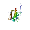
| ||||||||
|---|---|---|---|---|---|---|---|---|---|
| 1 |
| ||||||||
| Unit cell |
| ||||||||
| Components on special symmetry positions |
|
-Components
| #1: Protein | Mass: 15184.287 Da / Num. of mol.: 1 Source method: isolated from a genetically manipulated source Source: (gene. exp.) Chaetomium thermophilum (strain DSM 1495 / CBS 144.50 / IMI 039719) (fungus)Strain: DSM 1495 / CBS 144.50 / IMI 039719 / Gene: CTHT_0001260 / Production host:  | ||||||
|---|---|---|---|---|---|---|---|
| #2: Chemical |   Mass: 132.905 Da / Num. of mol.: 2 / Source method: obtained synthetically / Formula: Cs / Feature type: SUBJECT OF INVESTIGATION Mass: 132.905 Da / Num. of mol.: 2 / Source method: obtained synthetically / Formula: Cs / Feature type: SUBJECT OF INVESTIGATION#3: Chemical | ChemComp-CL / |   Mass: 35.453 Da / Num. of mol.: 1 / Source method: obtained synthetically / Formula: Cl Mass: 35.453 Da / Num. of mol.: 1 / Source method: obtained synthetically / Formula: Cl#4: Water | ChemComp-HOH / |  Mass: 18.015 Da / Num. of mol.: 96 / Source method: isolated from a natural source / Formula: H2O Mass: 18.015 Da / Num. of mol.: 96 / Source method: isolated from a natural source / Formula: H2OHas ligand of interest | Y | |
-Experimental details
-Experiment
| Experiment | Method: X-RAY DIFFRACTION / Number of used crystals: 1 |
|---|
- Sample preparation
| Crystal | Density Matthews: 2.51 Å3/Da / Density % sol: 51.06 % |
|---|---|
| Crystal grow | Temperature: 293.15 K / Method: vapor diffusion, hanging drop / Details: 0.6 M potassium chloride, 15 % (w/v) PEG 4000 |
-Data collection
| Diffraction | Mean temperature: 100 K / Serial crystal experiment: N | ||||||||||||||||||||||||||||||
|---|---|---|---|---|---|---|---|---|---|---|---|---|---|---|---|---|---|---|---|---|---|---|---|---|---|---|---|---|---|---|---|
| Diffraction source | Source: SYNCHROTRON / Site: ESRF  / Beamline: ID29 / Wavelength: 1.7712 Å / Beamline: ID29 / Wavelength: 1.7712 Å | ||||||||||||||||||||||||||||||
| Detector | Type: DECTRIS PILATUS 6M / Detector: PIXEL / Date: Oct 30, 2016 | ||||||||||||||||||||||||||||||
| Radiation | Protocol: SINGLE WAVELENGTH / Monochromatic (M) / Laue (L): M / Scattering type: x-ray | ||||||||||||||||||||||||||||||
| Radiation wavelength | Wavelength: 1.7712 Å / Relative weight: 1 | ||||||||||||||||||||||||||||||
| Reflection | Resolution: 1.9→42.27 Å / Num. obs: 12135 / % possible obs: 97.8 % / Redundancy: 23.7 % / CC1/2: 0.999 / Rmerge(I) obs: 0.093 / Rpim(I) all: 0.019 / Rrim(I) all: 0.095 / Net I/σ(I): 21.6 | ||||||||||||||||||||||||||||||
| Reflection shell | Diffraction-ID: 1
|
- Processing
| Software |
| ||||||||||||||||||||||||||||||||||||||||||||||||||||||||||||
|---|---|---|---|---|---|---|---|---|---|---|---|---|---|---|---|---|---|---|---|---|---|---|---|---|---|---|---|---|---|---|---|---|---|---|---|---|---|---|---|---|---|---|---|---|---|---|---|---|---|---|---|---|---|---|---|---|---|---|---|---|---|
| Refinement | Method to determine structure: SAD / Resolution: 1.9→42.27 Å / Cor.coef. Fo:Fc: 0.961 / Cor.coef. Fo:Fc free: 0.953 / SU B: 3.553 / SU ML: 0.101 / Cross valid method: THROUGHOUT / σ(F): 0 / ESU R: 0.131 / ESU R Free: 0.124 / Stereochemistry target values: MAXIMUM LIKELIHOOD Details: HYDROGENS HAVE BEEN ADDED IN THE RIDING POSITIONS U VALUES : REFINED INDIVIDUALLY
| ||||||||||||||||||||||||||||||||||||||||||||||||||||||||||||
| Solvent computation | Ion probe radii: 0.8 Å / Shrinkage radii: 0.8 Å / VDW probe radii: 1.2 Å / Solvent model: MASK | ||||||||||||||||||||||||||||||||||||||||||||||||||||||||||||
| Displacement parameters | Biso max: 117.18 Å2 / Biso mean: 43.584 Å2 / Biso min: 24.07 Å2
| ||||||||||||||||||||||||||||||||||||||||||||||||||||||||||||
| Refinement step | Cycle: final / Resolution: 1.9→42.27 Å
| ||||||||||||||||||||||||||||||||||||||||||||||||||||||||||||
| Refine LS restraints |
| ||||||||||||||||||||||||||||||||||||||||||||||||||||||||||||
| LS refinement shell | Resolution: 1.9→1.949 Å / Rfactor Rfree error: 0 / Total num. of bins used: 20
|
