 Movie
Movie Controller


+ Open data

- Basic information
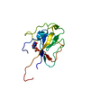
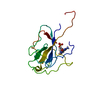
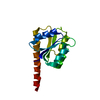
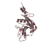
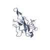
| Entry | Database: PDB / ID: 4lmc | ||||||
|---|---|---|---|---|---|---|---|
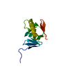
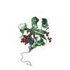
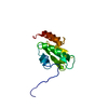
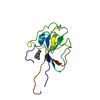
| Title | Crystal structure of HCoV-OC43 N-NTD complexed with CMP | ||||||
 Components Components | Nucleoprotein | ||||||
 Keywords Keywords | RNA BINDING PROTEIN / HCoV-OC43 / Nucleocapsid protein / N-terminal domain / RNA binding | ||||||
| Function / homology |  Function and homology information Function and homology informationhost cell / viral nucleocapsid / host cell endoplasmic reticulum-Golgi intermediate compartment / host cell Golgi apparatus / ribonucleoprotein complex / RNA binding Similarity search - Function | ||||||
| Biological species |  Human coronavirus Human coronavirus | ||||||
| Method |  X-RAY DIFFRACTION / SYNCHROTRON / MOLECULAR REPLACEMENT / Resolution: 1.742 Å X-RAY DIFFRACTION / SYNCHROTRON / MOLECULAR REPLACEMENT / Resolution: 1.742 Å | ||||||
 Authors Authors | Lin, S.Y. / Liu, C.L. / Hou, M.H. | ||||||
 Citation Citation | Journal: J.Med.Chem. / Year: 2014 Title: Structural basis for the identification of the N-terminal domain of coronavirus nucleocapsid protein as an antiviral target Authors: Lin, S.Y. / Liu, C.L. / Chang, Y.M. / Zhao, J. / Perlman, S. / Hou, M.H. | ||||||
| History |
|
- Structure visualization
| Structure viewer | Molecule: MolmilJmol/JSmol |
|---|
- Downloads & links
-Download
| PDBx/mmCIF format | 4lmc.cif.gz | 64.8 KB | Display | PDBx/mmCIF format |
|---|---|---|---|---|
| PDB format | pdb4lmc.ent.gz | 48.2 KB | Display | PDB format |
| PDBx/mmJSON format | 4lmc.json.gz | Tree view | PDBx/mmJSON format | |
| Others |  Other downloads Other downloads |
-Validation report
| Arichive directory | https://data.pdbj.org/pub/pdb/validation_reports/lm/4lmcftp://data.pdbj.org/pub/pdb/validation_reports/lm/4lmc | HTTPS FTP |
|---|
-Related structure data
| Related structure data |  4kxjC  4li4C  4lm7C  4lm9C  4j3kS C: citing same article ( S: Starting model for refinement |
|---|---|
| Similar structure data |


-Links
 PDBj
PDBj- Assembly
| Deposited unit | 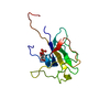
| ||||||||
|---|---|---|---|---|---|---|---|---|---|
| 1 |
| ||||||||
| Unit cell |
|
-Components
| #1: Protein | Mass: 15350.822 Da / Num. of mol.: 1 / Fragment: UNP residues 55-187 Source method: isolated from a genetically manipulated source Source: (gene. exp.) Human coronavirus / Strain: OC43 / Gene: N / Production host:  |
|---|---|
| #2: Chemical | ChemComp-C5P /   Mass: 323.197 Da / Num. of mol.: 1 / Source method: obtained synthetically / Formula: C9H14N3O8P Mass: 323.197 Da / Num. of mol.: 1 / Source method: obtained synthetically / Formula: C9H14N3O8P |
| #3: Water | ChemComp-HOH /  Mass: 18.015 Da / Num. of mol.: 118 / Source method: isolated from a natural source / Formula: H2O Mass: 18.015 Da / Num. of mol.: 118 / Source method: isolated from a natural source / Formula: H2O |
-Experimental details
-Experiment
| Experiment | Method: X-RAY DIFFRACTION / Number of used crystals: 1 |
|---|
- Sample preparation
| Crystal | Density Matthews: 2.7 Å3/Da / Density % sol: 54.52 % |
|---|---|
| Crystal grow | Temperature: 293 K / Method: vapor diffusion, sitting drop / pH: 7.5 Details: 25% PEG1500, 0.25M SPG, pH 7.5, VAPOR DIFFUSION, SITTING DROP, temperature 293K |
-Data collection
| Diffraction | Mean temperature: 100 K |
|---|---|
| Diffraction source | Source: SYNCHROTRON / Site: NSRRC  / Beamline: BL13B1 / Wavelength: 1 Å / Beamline: BL13B1 / Wavelength: 1 Å |
| Detector | Type: ADSC QUANTUM 315r / Detector: CCD / Date: Dec 12, 2011 |
| Radiation | Monochromator: LN2-Cooled, Fixed-Exit Double Crystal Monochromator Protocol: SINGLE WAVELENGTH / Monochromatic (M) / Laue (L): M / Scattering type: x-ray |
| Radiation wavelength | Wavelength: 1 Å / Relative weight: 1 |
| Reflection | Resolution: 1.74→30 Å / Num. all: 16152 / Num. obs: 16152 / % possible obs: 94.8 % / Observed criterion σ(F): 0 / Observed criterion σ(I): -3 / Biso Wilson estimate: 24.72 Å2 |
| Reflection shell | Resolution: 1.74→1.8 Å / % possible all: 94.8 |
- Processing
| Software | Name: PHENIX / Version: (phenix.refine: 1.8.1_1168) / Classification: refinement | |||||||||||||||||||||||||||||||||||||||||||||||||||||||||||||||||||||||||||||||||||||||||||
|---|---|---|---|---|---|---|---|---|---|---|---|---|---|---|---|---|---|---|---|---|---|---|---|---|---|---|---|---|---|---|---|---|---|---|---|---|---|---|---|---|---|---|---|---|---|---|---|---|---|---|---|---|---|---|---|---|---|---|---|---|---|---|---|---|---|---|---|---|---|---|---|---|---|---|---|---|---|---|---|---|---|---|---|---|---|---|---|---|---|---|---|---|
| Refinement | Method to determine structure: MOLECULAR REPLACEMENT Starting model: 4J3K Resolution: 1.742→26.807 Å / Occupancy max: 1 / Occupancy min: 1 / FOM work R set: 0.8157 / SU ML: 0.21 / σ(F): 1.37 / Phase error: 25.42 / Stereochemistry target values: Engh & Huber
| |||||||||||||||||||||||||||||||||||||||||||||||||||||||||||||||||||||||||||||||||||||||||||
| Solvent computation | Shrinkage radii: 0.9 Å / VDW probe radii: 1.11 Å / Solvent model: FLAT BULK SOLVENT MODEL | |||||||||||||||||||||||||||||||||||||||||||||||||||||||||||||||||||||||||||||||||||||||||||
| Displacement parameters | Biso max: 85.81 Å2 / Biso mean: 33.1738 Å2 / Biso min: 11.27 Å2 | |||||||||||||||||||||||||||||||||||||||||||||||||||||||||||||||||||||||||||||||||||||||||||
| Refinement step | Cycle: LAST / Resolution: 1.742→26.807 Å
| |||||||||||||||||||||||||||||||||||||||||||||||||||||||||||||||||||||||||||||||||||||||||||
| Refine LS restraints |
| |||||||||||||||||||||||||||||||||||||||||||||||||||||||||||||||||||||||||||||||||||||||||||
| LS refinement shell | Refine-ID: X-RAY DIFFRACTION / Total num. of bins used: 12
|
