 Movie
Movie Controller

[English] 日本語
 Yorodumi
- PDB-7bmh: Crystal structure of a light-driven proton pump LR (Mac) from Lep... -
+ Open data

- Basic information
| Entry | Database: PDB / ID: 7bmh | |||||||||||||||
|---|---|---|---|---|---|---|---|---|---|---|---|---|---|---|---|---|
| Title | Crystal structure of a light-driven proton pump LR (Mac) from Leptosphaeria maculans | |||||||||||||||
 Components Components | Opsin | |||||||||||||||
 Keywords Keywords | MEMBRANE PROTEIN / rhodopsin / bacteriorhodopsin / retinal / optogenetics / proton pump / photocycle / eukaryotic rhodopsin / ion transport | |||||||||||||||
| Function / homology |  Function and homology information Function and homology informationmonoatomic ion channel activity / photoreceptor activity / phototransduction / membrane Similarity search - Function | |||||||||||||||
| Biological species |  Leptosphaeria maculans (blackleg of rapeseed fungus) Leptosphaeria maculans (blackleg of rapeseed fungus) | |||||||||||||||
| Method |  X-RAY DIFFRACTION / SYNCHROTRON / MOLECULAR REPLACEMENT / Resolution: 2.2 Å X-RAY DIFFRACTION / SYNCHROTRON / MOLECULAR REPLACEMENT / Resolution: 2.2 Å | |||||||||||||||
 Authors Authors | Kovalev, K. / Zabelskii, D. / Dmitrieva, N. / Volkov, O. / Shevchenko, V. / Astashkin, R. / Zinovev, E. / Gordeliy, V. | |||||||||||||||
| Funding support |  Russian Federation, Russian Federation,  France, 4items France, 4items
| |||||||||||||||
 Citation Citation | Journal: Commun Biol / Year: 2021 Title: Structure-based insights into evolution of rhodopsins. Authors: Zabelskii, D. / Dmitrieva, N. / Volkov, O. / Shevchenko, V. / Kovalev, K. / Balandin, T. / Soloviov, D. / Astashkin, R. / Zinovev, E. / Alekseev, A. / Round, E. / Polovinkin, V. / Chizhov, I. ...Authors: Zabelskii, D. / Dmitrieva, N. / Volkov, O. / Shevchenko, V. / Kovalev, K. / Balandin, T. / Soloviov, D. / Astashkin, R. / Zinovev, E. / Alekseev, A. / Round, E. / Polovinkin, V. / Chizhov, I. / Rogachev, A. / Okhrimenko, I. / Borshchevskiy, V. / Chupin, V. / Buldt, G. / Yutin, N. / Bamberg, E. / Koonin, E. / Gordeliy, V. | |||||||||||||||
| History |
|
- Structure visualization
| Structure viewer | Molecule: MolmilJmol/JSmol |
|---|
- Downloads & links
-Download
| PDBx/mmCIF format | 7bmh.cif.gz | 125.6 KB | Display | PDBx/mmCIF format |
|---|---|---|---|---|
| PDB format | pdb7bmh.ent.gz | 92.4 KB | Display | PDB format |
| PDBx/mmJSON format | 7bmh.json.gz | Tree view | PDBx/mmJSON format | |
| Others |  Other downloads Other downloads |
-Validation report
| Summary document | 7bmh_validation.pdf.gz | 943.7 KB | Display | wwPDB validaton report |
|---|---|---|---|---|
| Full document | 7bmh_full_validation.pdf.gz | 886 KB | Display | |
| Data in XML | 7bmh_validation.xml.gz | 23.6 KB | Display | |
| Data in CIF | 7bmh_validation.cif.gz | 31.7 KB | Display | |
| Arichive directory | https://data.pdbj.org/pub/pdb/validation_reports/bm/7bmhftp://data.pdbj.org/pub/pdb/validation_reports/bm/7bmh | HTTPS FTP |
-Related structure data
| Related structure data | 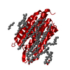 1c3wS S: Starting model for refinement |
|---|---|
| Similar structure data |
-Links
 PDBj
PDBj


- Assembly
| Deposited unit | 
| ||||||||
|---|---|---|---|---|---|---|---|---|---|
| 1 | 
| ||||||||
| 2 | 
| ||||||||
| Unit cell |
|
-Components
| #1: Protein | Mass: 35970.625 Da / Num. of mol.: 2 Source method: isolated from a genetically manipulated source Source: (gene. exp.) Leptosphaeria maculans (blackleg of rapeseed fungus)Gene: ops / Production host:  Leishmania tarentolae (eukaryote) / References: UniProt: Q9HGT7 Leishmania tarentolae (eukaryote) / References: UniProt: Q9HGT7#2: Chemical | ChemComp-LFA /   Mass: 282.547 Da / Num. of mol.: 43 / Source method: obtained synthetically / Formula: C20H42 Mass: 282.547 Da / Num. of mol.: 43 / Source method: obtained synthetically / Formula: C20H42#3: Chemical | ChemComp-OLA /   Mass: 282.461 Da / Num. of mol.: 6 / Source method: obtained synthetically / Formula: C18H34O2 Mass: 282.461 Da / Num. of mol.: 6 / Source method: obtained synthetically / Formula: C18H34O2#4: Water | ChemComp-HOH / |  Mass: 18.015 Da / Num. of mol.: 102 / Source method: isolated from a natural source / Formula: H2O Mass: 18.015 Da / Num. of mol.: 102 / Source method: isolated from a natural source / Formula: H2OHas ligand of interest | Y | |
|---|
-Experimental details
-Experiment
| Experiment | Method: X-RAY DIFFRACTION / Number of used crystals: 1 |
|---|
- Sample preparation
| Crystal | Density Matthews: 2.31 Å3/Da / Density % sol: 46.83 % |
|---|---|
| Crystal grow | Temperature: 293 K / Method: lipidic cubic phase Details: 100 mM HEPES pH 7.0, 4% PEG 3350, 1 M sodium malonate pH 7.0 |
-Data collection
| Diffraction | Mean temperature: 100 K / Serial crystal experiment: N | ||||||||||||||||||||||||||||||
|---|---|---|---|---|---|---|---|---|---|---|---|---|---|---|---|---|---|---|---|---|---|---|---|---|---|---|---|---|---|---|---|
| Diffraction source | Source: SYNCHROTRON / Site: ESRF / Beamline: ID30B / Wavelength: 0.97625 Å | ||||||||||||||||||||||||||||||
| Detector | Type: DECTRIS PILATUS 6M-F / Detector: PIXEL / Date: Aug 28, 2018 | ||||||||||||||||||||||||||||||
| Radiation | Protocol: SINGLE WAVELENGTH / Monochromatic (M) / Laue (L): M / Scattering type: x-ray | ||||||||||||||||||||||||||||||
| Radiation wavelength | Wavelength: 0.97625 Å / Relative weight: 1 | ||||||||||||||||||||||||||||||
| Reflection | Resolution: 2.2→49.39 Å / Num. obs: 34762 / % possible obs: 99.8 % / Redundancy: 4.8 % / Biso Wilson estimate: 31.58 Å2 / CC1/2: 0.996 / Rmerge(I) obs: 0.172 / Rpim(I) all: 0.086 / Rrim(I) all: 0.193 / Net I/σ(I): 5.7 / Num. measured all: 168155 / Scaling rejects: 1 | ||||||||||||||||||||||||||||||
| Reflection shell | Diffraction-ID: 1
|
- Processing
| Software |
| |||||||||||||||||||||||||||||||||||||||||||||||||||||||||||||||||||||||||||||||||||||||||||
|---|---|---|---|---|---|---|---|---|---|---|---|---|---|---|---|---|---|---|---|---|---|---|---|---|---|---|---|---|---|---|---|---|---|---|---|---|---|---|---|---|---|---|---|---|---|---|---|---|---|---|---|---|---|---|---|---|---|---|---|---|---|---|---|---|---|---|---|---|---|---|---|---|---|---|---|---|---|---|---|---|---|---|---|---|---|---|---|---|---|---|---|---|
| Refinement | Method to determine structure: MOLECULAR REPLACEMENT Starting model: 1C3W Resolution: 2.2→19.92 Å / SU ML: 0.31 / Cross valid method: THROUGHOUT / σ(F): 1.34 / Phase error: 35.33 / Stereochemistry target values: ML
| |||||||||||||||||||||||||||||||||||||||||||||||||||||||||||||||||||||||||||||||||||||||||||
| Solvent computation | Shrinkage radii: 0.9 Å / VDW probe radii: 1.11 Å / Solvent model: FLAT BULK SOLVENT MODEL | |||||||||||||||||||||||||||||||||||||||||||||||||||||||||||||||||||||||||||||||||||||||||||
| Displacement parameters | Biso max: 118.05 Å2 / Biso mean: 36.1578 Å2 / Biso min: 17.23 Å2 | |||||||||||||||||||||||||||||||||||||||||||||||||||||||||||||||||||||||||||||||||||||||||||
| Refinement step | Cycle: final / Resolution: 2.2→19.92 Å
| |||||||||||||||||||||||||||||||||||||||||||||||||||||||||||||||||||||||||||||||||||||||||||
| LS refinement shell | Refine-ID: X-RAY DIFFRACTION / Rfactor Rfree error: 0 / Total num. of bins used: 12
|
