 Movie
Movie Controller


+ Open data

- Basic information
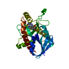
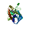
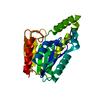
| Entry | Database: PDB / ID: 7bk8 | ||||||||||||
|---|---|---|---|---|---|---|---|---|---|---|---|---|---|
| Title | X-ray crystal structure of Pseudomonas aeruginosa MagC | ||||||||||||
 Components Components | MagC | ||||||||||||
 Keywords Keywords | HYDROLASE / alpha-2-macroglobulin / peptidoglycan / NlpC/P60 / endopeptidase / MagC | ||||||||||||
| Function / homology | Protein of unknown function DUF1175 / Protein of unknown function (DUF1175) / PHOSPHATE ION / Uncharacterized protein PA4490 Function and homology information Function and homology information | ||||||||||||
| Biological species |   Pseudomonas aeruginosa (bacteria) Pseudomonas aeruginosa (bacteria) | ||||||||||||
| Method |  X-RAY DIFFRACTION / SYNCHROTRON / AB INITIO PHASING / Resolution: 1.74 Å X-RAY DIFFRACTION / SYNCHROTRON / AB INITIO PHASING / Resolution: 1.74 Å | ||||||||||||
 Authors Authors | Zouhir, S. / Contreras-Martel, C. / Maragno Trindade, D. / Attree, I. / Dessen, A. / Macheboeuf, P. | ||||||||||||
| Funding support |  Brazil, Brazil,  France, 3items France, 3items
| ||||||||||||
 Citation Citation | Journal: Febs Lett. / Year: 2021 Title: MagC is a NplC/P60-like member of the alpha-2-macroglobulin Mag complex of Pseudomonas aeruginosa that interacts with peptidoglycan. Authors: Zouhir, S. / Contreras-Martel, C. / Maragno Trindade, D. / Attree, I. / Dessen, A. / Macheboeuf, P. | ||||||||||||
| History |
|
- Structure visualization
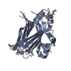
| Structure viewer | Molecule: MolmilJmol/JSmol |
|---|
- Downloads & links
-Download
| PDBx/mmCIF format | 7bk8.cif.gz | 89.1 KB | Display | PDBx/mmCIF format |
|---|---|---|---|---|
| PDB format | pdb7bk8.ent.gz | 64.8 KB | Display | PDB format |
| PDBx/mmJSON format | 7bk8.json.gz | Tree view | PDBx/mmJSON format | |
| Others |  Other downloads Other downloads |
-Validation report
| Summary document | 7bk8_validation.pdf.gz | 429.3 KB | Display | wwPDB validaton report |
|---|---|---|---|---|
| Full document | 7bk8_full_validation.pdf.gz | 431 KB | Display | |
| Data in XML | 7bk8_validation.xml.gz | 10 KB | Display | |
| Data in CIF | 7bk8_validation.cif.gz | 13.1 KB | Display | |
| Arichive directory | https://data.pdbj.org/pub/pdb/validation_reports/bk/7bk8ftp://data.pdbj.org/pub/pdb/validation_reports/bk/7bk8 | HTTPS FTP |
-Related structure data
| Similar structure data |
|---|


-Links
 PDBj
PDBj- Assembly
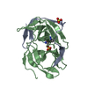
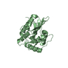
| Deposited unit | 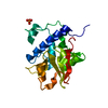
| |||||||||
|---|---|---|---|---|---|---|---|---|---|---|
| 1 |
| |||||||||
| Unit cell |
| |||||||||
| Components on special symmetry positions |
|
-Components
| #1: Protein | Mass: 24028.264 Da / Num. of mol.: 1 Source method: isolated from a genetically manipulated source Details: N-terminal signal peptide was replaced by a hexa-histidine tag Source: (gene. exp.) Pseudomonas aeruginosa (strain ATCC 15692 / DSM 22644 / CIP 104116 / JCM 14847 / LMG 12228 / 1C / PRS 101 / PAO1) (bacteria)Strain: ATCC 15692 / DSM 22644 / CIP 104116 / JCM 14847 / LMG 12228 / 1C / PRS 101 / PAO1 Gene: PA4490 / Plasmid: pET28a (Novagen) / Production host: |
|---|---|
| #2: Chemical | ChemComp-PO4 /   Mass: 94.971 Da / Num. of mol.: 1 / Source method: obtained synthetically / Formula: PO4 Mass: 94.971 Da / Num. of mol.: 1 / Source method: obtained synthetically / Formula: PO4 |
| #3: Water | ChemComp-HOH /  Mass: 18.015 Da / Num. of mol.: 82 / Source method: isolated from a natural source / Formula: H2O Mass: 18.015 Da / Num. of mol.: 82 / Source method: isolated from a natural source / Formula: H2O |
| Has ligand of interest | N |
-Experimental details
-Experiment
| Experiment | Method: X-RAY DIFFRACTION / Number of used crystals: 1 |
|---|
- Sample preparation
| Crystal | Density Matthews: 2.02 Å3/Da / Density % sol: 39.12 % / Description: Tiny baguettes |
|---|---|
| Crystal grow | Temperature: 293 K / Method: vapor diffusion / pH: 6.5 / Details: 30% w/v PEG6000, 100 mM Citrate pH 6.5 |
-Data collection
| Diffraction | Mean temperature: 100 K / Serial crystal experiment: N |
|---|---|
| Diffraction source | Source: SYNCHROTRON / Site: LNLS / Beamline: W01B-MX2 / Wavelength: 1.45868 Å |
| Detector | Type: DECTRIS PILATUS 2M / Detector: PIXEL / Date: Jun 29, 2016 |
| Radiation | Protocol: SINGLE WAVELENGTH / Monochromatic (M) / Laue (L): M / Scattering type: x-ray |
| Radiation wavelength | Wavelength: 1.45868 Å / Relative weight: 1 |
| Reflection | Resolution: 1.74→37.79 Å / Num. obs: 18977 / % possible obs: 98.4 % / Observed criterion σ(I): -3 / Redundancy: 6.6 % / Biso Wilson estimate: 31.783 Å2 / CC1/2: 1 / Rsym value: 0.045 / Net I/σ(I): 26.91 |
| Reflection shell | Resolution: 1.74→1.85 Å / Redundancy: 4.9 % / Mean I/σ(I) obs: 3 / Num. unique obs: 3021 / CC1/2: 0.889 / Rsym value: 0.569 / % possible all: 93.7 |
- Processing
| Software |
| |||||||||||||||||||||||||||||||||||||||||||||||||||||||||||||||||||||||||||||||||||||||||||||||||||||||||||||||||||||||||||||||||||||||||||||||||||||||||||||||||||||||||||||||||||||||||||||||||||||||||||||||||||||||||||||||||||||||
|---|---|---|---|---|---|---|---|---|---|---|---|---|---|---|---|---|---|---|---|---|---|---|---|---|---|---|---|---|---|---|---|---|---|---|---|---|---|---|---|---|---|---|---|---|---|---|---|---|---|---|---|---|---|---|---|---|---|---|---|---|---|---|---|---|---|---|---|---|---|---|---|---|---|---|---|---|---|---|---|---|---|---|---|---|---|---|---|---|---|---|---|---|---|---|---|---|---|---|---|---|---|---|---|---|---|---|---|---|---|---|---|---|---|---|---|---|---|---|---|---|---|---|---|---|---|---|---|---|---|---|---|---|---|---|---|---|---|---|---|---|---|---|---|---|---|---|---|---|---|---|---|---|---|---|---|---|---|---|---|---|---|---|---|---|---|---|---|---|---|---|---|---|---|---|---|---|---|---|---|---|---|---|---|---|---|---|---|---|---|---|---|---|---|---|---|---|---|---|---|---|---|---|---|---|---|---|---|---|---|---|---|---|---|---|---|---|---|---|---|---|---|---|---|---|---|---|---|---|---|---|---|---|
| Refinement | Method to determine structure: AB INITIO PHASING / Resolution: 1.74→37.785 Å / Cor.coef. Fo:Fc: 0.959 / Cor.coef. Fo:Fc free: 0.947 / SU B: 6.076 / SU ML: 0.093 / Cross valid method: FREE R-VALUE / ESU R: 0.137 / ESU R Free: 0.124 Details: Hydrogens have been added in their riding positions
| |||||||||||||||||||||||||||||||||||||||||||||||||||||||||||||||||||||||||||||||||||||||||||||||||||||||||||||||||||||||||||||||||||||||||||||||||||||||||||||||||||||||||||||||||||||||||||||||||||||||||||||||||||||||||||||||||||||||
| Solvent computation | Ion probe radii: 0.8 Å / Shrinkage radii: 0.8 Å / VDW probe radii: 1.2 Å / Solvent model: BABINET MODEL PLUS MASK | |||||||||||||||||||||||||||||||||||||||||||||||||||||||||||||||||||||||||||||||||||||||||||||||||||||||||||||||||||||||||||||||||||||||||||||||||||||||||||||||||||||||||||||||||||||||||||||||||||||||||||||||||||||||||||||||||||||||
| Displacement parameters | Biso mean: 39.74 Å2
| |||||||||||||||||||||||||||||||||||||||||||||||||||||||||||||||||||||||||||||||||||||||||||||||||||||||||||||||||||||||||||||||||||||||||||||||||||||||||||||||||||||||||||||||||||||||||||||||||||||||||||||||||||||||||||||||||||||||
| Refinement step | Cycle: LAST / Resolution: 1.74→37.785 Å
| |||||||||||||||||||||||||||||||||||||||||||||||||||||||||||||||||||||||||||||||||||||||||||||||||||||||||||||||||||||||||||||||||||||||||||||||||||||||||||||||||||||||||||||||||||||||||||||||||||||||||||||||||||||||||||||||||||||||
| Refine LS restraints |
| |||||||||||||||||||||||||||||||||||||||||||||||||||||||||||||||||||||||||||||||||||||||||||||||||||||||||||||||||||||||||||||||||||||||||||||||||||||||||||||||||||||||||||||||||||||||||||||||||||||||||||||||||||||||||||||||||||||||
| LS refinement shell | Refine-ID: X-RAY DIFFRACTION / Total num. of bins used: 20
| |||||||||||||||||||||||||||||||||||||||||||||||||||||||||||||||||||||||||||||||||||||||||||||||||||||||||||||||||||||||||||||||||||||||||||||||||||||||||||||||||||||||||||||||||||||||||||||||||||||||||||||||||||||||||||||||||||||||
| Refinement TLS params. | Method: refined / Refine-ID: X-RAY DIFFRACTION
| |||||||||||||||||||||||||||||||||||||||||||||||||||||||||||||||||||||||||||||||||||||||||||||||||||||||||||||||||||||||||||||||||||||||||||||||||||||||||||||||||||||||||||||||||||||||||||||||||||||||||||||||||||||||||||||||||||||||
| Refinement TLS group |
|
