 Movie
Movie Controller


+ Open data

- Basic information
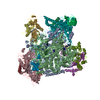
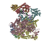
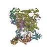
| Entry | Database: PDB / ID: 7bef | ||||||||||||
|---|---|---|---|---|---|---|---|---|---|---|---|---|---|
| Title | Structures of class II bacterial transcription complexes | ||||||||||||
 Components Components |
| ||||||||||||
 Keywords Keywords | TRANSCRIPTION / antibiotic resistance / bacterial transcription activator / cryoEM | ||||||||||||
| Function / homology |  Function and homology information Function and homology informationsigma factor antagonist complex / RNA polymerase complex / submerged biofilm formation / cellular response to cell envelope stress / regulation of DNA-templated transcription initiation / sigma factor activity / bacterial-type flagellum assembly / bacterial-type RNA polymerase core enzyme binding / cytosolic DNA-directed RNA polymerase complex / bacterial-type flagellum-dependent cell motility ...sigma factor antagonist complex / RNA polymerase complex / submerged biofilm formation / cellular response to cell envelope stress / regulation of DNA-templated transcription initiation / sigma factor activity / bacterial-type flagellum assembly / bacterial-type RNA polymerase core enzyme binding / cytosolic DNA-directed RNA polymerase complex / bacterial-type flagellum-dependent cell motility / nitrate assimilation / regulation of DNA-templated transcription elongation / transcription elongation factor complex / DNA-directed RNA polymerase complex / transcription antitermination / cell motility / DNA-templated transcription initiation / ribonucleoside binding / DNA-directed RNA polymerase / DNA-directed RNA polymerase activity / response to heat / protein-containing complex assembly / sequence-specific DNA binding / intracellular iron ion homeostasis / protein dimerization activity / transcription cis-regulatory region binding / DNA-binding transcription factor activity / response to antibiotic / negative regulation of DNA-templated transcription / regulation of DNA-templated transcription / DNA-templated transcription / magnesium ion binding / DNA binding / zinc ion binding / membrane / cytoplasm / cytosol Similarity search - Function | ||||||||||||
| Biological species |  Klebsiella pneumoniae (bacteria) Klebsiella pneumoniae (bacteria) | ||||||||||||
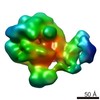
| Method | ELECTRON MICROSCOPY / single particle reconstruction / cryo EM / Resolution: 4.5 Å | ||||||||||||
 Authors Authors | Hao, M. / Ye, F.Z. / Zhang, X.D. | ||||||||||||
| Funding support |  United Kingdom, United Kingdom,  China, 3items China, 3items
| ||||||||||||
 Citation Citation | Journal: Adv Sci (Weinh) / Year: 2022 Title: Structures of Class I and Class II Transcription Complexes Reveal the Molecular Basis of RamA-Dependent Transcription Activation. Authors: Min Hao / Fuzhou Ye / Milija Jovanovic / Ioly Kotta-Loizou / Qingqing Xu / Xiaohua Qin / Martin Buck / Xiaodong Zhang / Minggui Wang / Abstract: Transcription activator RamA is linked to multidrug resistance of Klebsiella pneumoniae through controlling genes that encode efflux pumps (acrA) and porin-regulating antisense RNA (micF). In ...Transcription activator RamA is linked to multidrug resistance of Klebsiella pneumoniae through controlling genes that encode efflux pumps (acrA) and porin-regulating antisense RNA (micF). In bacteria, σ , together with activators, controls the majority of genes by recruiting RNA polymerase (RNAP) to the promoter regions. RNAP and σ form a holoenzyme that recognizes -35 and -10 promoter DNA consensus sites. Many activators bind upstream from the holoenzyme and can be broadly divided into two classes. RamA acts as a class I activator on acrA and class II activator on micF, respectively. The authors present biochemical and structural data on RamA in complex with RNAP-σ at the two promoters and the data reveal the molecular basis for how RamA assembles and interacts with core RNAP and activates transcription that contributes to antibiotic resistance. Further, comparing with CAP/TAP complexes reveals common and activator-specific features in activator binding and uncovers distinct roles of the two C-terminal domains of RNAP α subunit. | ||||||||||||
| History |
|
- Structure visualization
| Movie |
Movie viewer |
|---|---|
| Structure viewer | Molecule: MolmilJmol/JSmol |
- Downloads & links
-Download
| PDBx/mmCIF format | 7bef.cif.gz | 760.9 KB | Display | PDBx/mmCIF format |
|---|---|---|---|---|
| PDB format | pdb7bef.ent.gz | 611.2 KB | Display | PDB format |
| PDBx/mmJSON format | 7bef.json.gz | Tree view | PDBx/mmJSON format | |
| Others |  Other downloads Other downloads |
-Validation report
| Arichive directory | https://data.pdbj.org/pub/pdb/validation_reports/be/7befftp://data.pdbj.org/pub/pdb/validation_reports/be/7bef | HTTPS FTP |
|---|
-Related structure data
| Related structure data | 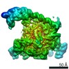 12156MC  7begC M: map data used to model this data C: citing same article ( |
|---|---|
| Similar structure data |


-Links
 PDBj
PDBj


- Assembly
| Deposited unit | 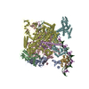
|
|---|---|
| 1 |
|
-Components
-DNA-directed RNA polymerase subunit ... , 4 types, 5 molecules ABCDE
| #1: Protein | Mass: 36558.680 Da / Num. of mol.: 2 Source method: isolated from a genetically manipulated source Source: (gene. exp.) Strain: K12 / Gene: rpoA, pez, phs, sez, b3295, JW3257 / Production host: #2: Protein | | Mass: 150820.875 Da / Num. of mol.: 1 Source method: isolated from a genetically manipulated source Source: (gene. exp.) Strain: K12 Gene: rpoB, groN, nitB, rif, ron, stl, stv, tabD, b3987, JW3950 Production host: #3: Protein | | Mass: 155366.781 Da / Num. of mol.: 1 Source method: isolated from a genetically manipulated source Source: (gene. exp.) Strain: K12 / Gene: rpoC, tabB, b3988, JW3951 / Production host: #4: Protein | | Mass: 10249.547 Da / Num. of mol.: 1 Source method: isolated from a genetically manipulated source Source: (gene. exp.) Strain: K12 / Gene: rpoZ, b3649, JW3624 / Production host: |
|---|
-Protein , 2 types, 2 molecules FG
| #5: Protein | Mass: 72344.500 Da / Num. of mol.: 1 Source method: isolated from a genetically manipulated source Source: (gene. exp.) Strain: K12 / Gene: rpoD, alt, b3067, JW3039 / Production host: |
|---|---|
| #6: Protein | Mass: 15510.520 Da / Num. of mol.: 1 Source method: isolated from a genetically manipulated source Source: (gene. exp.) Klebsiella pneumoniae (bacteria) / Gene: ramA / Production host: |
-PmicF promoter ... , 2 types, 2 molecules TN
| #7: DNA chain | Mass: 22584.451 Da / Num. of mol.: 1 Source method: isolated from a genetically manipulated source Source: (gene. exp.) Klebsiella pneumoniae (bacteria) / Production host: |
|---|---|
| #8: DNA chain | Mass: 22480.471 Da / Num. of mol.: 1 Source method: isolated from a genetically manipulated source Source: (gene. exp.) Klebsiella pneumoniae (bacteria) / Production host: |
-Experimental details
-Experiment
| Experiment | Method: ELECTRON MICROSCOPY |
|---|---|
| EM experiment | Aggregation state: PARTICLE / 3D reconstruction method: single particle reconstruction |
- Sample preparation
| Component |
| ||||||||||||||||||||||||
|---|---|---|---|---|---|---|---|---|---|---|---|---|---|---|---|---|---|---|---|---|---|---|---|---|---|
| Molecular weight | Value: 0.56 MDa / Experimental value: NO | ||||||||||||||||||||||||
| Source (natural) |
| ||||||||||||||||||||||||
| Source (recombinant) |
| ||||||||||||||||||||||||
| Buffer solution | pH: 8 / Details: 20 mM Tris pH 8, 50 mM NaCl | ||||||||||||||||||||||||
| Buffer component |
| ||||||||||||||||||||||||
| Specimen | Conc.: 0.5 mg/ml / Embedding applied: NO / Shadowing applied: NO / Staining applied: NO / Vitrification applied: YES | ||||||||||||||||||||||||
| Specimen support | Grid material: COPPER / Grid mesh size: 200 divisions/in. / Grid type: Quantifoil | ||||||||||||||||||||||||
| Vitrification | Instrument: FEI VITROBOT MARK IV / Cryogen name: ETHANE / Humidity: 100 % / Chamber temperature: 100 K |
- Electron microscopy imaging
| Experimental equipment |  Model: Titan Krios / Image courtesy: FEI Company |
|---|---|
| Microscopy | Model: FEI TITAN KRIOS |
| Electron gun | Electron source:  FIELD EMISSION GUN / Accelerating voltage: 300 kV / Illumination mode: OTHER FIELD EMISSION GUN / Accelerating voltage: 300 kV / Illumination mode: OTHER |
| Electron lens | Mode: BRIGHT FIELD / Nominal magnification: 75000 X / Cs: 2.7 mm / C2 aperture diameter: 70 µm |
| Specimen holder | Specimen holder model: FEI TITAN KRIOS AUTOGRID HOLDER |
| Image recording | Electron dose: 79.1 e/Å2 / Film or detector model: FEI FALCON III (4k x 4k) / Num. of grids imaged: 1 / Num. of real images: 2878 |
- Processing
| EM software |
| ||||||||||||||||||||||||
|---|---|---|---|---|---|---|---|---|---|---|---|---|---|---|---|---|---|---|---|---|---|---|---|---|---|
| CTF correction | Type: PHASE FLIPPING AND AMPLITUDE CORRECTION | ||||||||||||||||||||||||
| Particle selection | Num. of particles selected: 774955 | ||||||||||||||||||||||||
| 3D reconstruction | Resolution: 4.5 Å / Resolution method: FSC 0.143 CUT-OFF / Num. of particles: 74282 / Algorithm: FOURIER SPACE / Num. of class averages: 1 / Symmetry type: POINT | ||||||||||||||||||||||||
| Atomic model building | Protocol: RIGID BODY FIT / Space: REAL | ||||||||||||||||||||||||
| Atomic model building | PDB-ID: 4YLP Accession code: 4YLP / Source name: PDB / Type: experimental model |

