Biotechnology and Biological Sciences Research Council (BBSRC)
BB/N007816/1
United Kingdom
Medical Research Council (MRC, United Kingdom)
MR/P007503/1
United Kingdom
National Science Foundation (NSF, China)
81991531
China
Citation
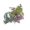
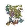
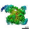
Journal: Adv Sci (Weinh) / Year: 2022 Title: Structures of Class I and Class II Transcription Complexes Reveal the Molecular Basis of RamA-Dependent Transcription Activation. Authors: Min Hao / Fuzhou Ye / Milija Jovanovic / Ioly Kotta-Loizou / Qingqing Xu / Xiaohua Qin / Martin Buck / Xiaodong Zhang / Minggui Wang / Abstract: Transcription activator RamA is linked to multidrug resistance of Klebsiella pneumoniae through controlling genes that encode efflux pumps (acrA) and porin-regulating antisense RNA (micF). In ...Transcription activator RamA is linked to multidrug resistance of Klebsiella pneumoniae through controlling genes that encode efflux pumps (acrA) and porin-regulating antisense RNA (micF). In bacteria, σ , together with activators, controls the majority of genes by recruiting RNA polymerase (RNAP) to the promoter regions. RNAP and σ form a holoenzyme that recognizes -35 and -10 promoter DNA consensus sites. Many activators bind upstream from the holoenzyme and can be broadly divided into two classes. RamA acts as a class I activator on acrA and class II activator on micF, respectively. The authors present biochemical and structural data on RamA in complex with RNAP-σ at the two promoters and the data reveal the molecular basis for how RamA assembles and interacts with core RNAP and activates transcription that contributes to antibiotic resistance. Further, comparing with CAP/TAP complexes reveals common and activator-specific features in activator binding and uncovers distinct roles of the two C-terminal domains of RNAP α subunit.
History
Deposition
Dec 23, 2020
-
Header (metadata) release
Dec 1, 2021
-
Map release
Dec 1, 2021
-
Update
Jul 10, 2024
-
Current status
Jul 10, 2024
Processing site: PDBe / Status: Released
-
Structure visualization
Movie
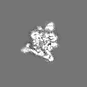
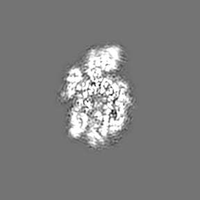
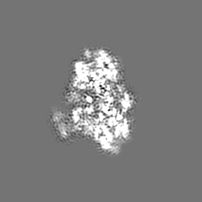
Surface view with section colored by density value
In the structure databanks used in Yorodumi, some data are registered as the other names, "COVID-19 virus" and "2019-nCoV". Here are the details of the virus and the list of structure data.
Jan 31, 2019. EMDB accession codes are about to change! (news from PDBe EMDB page)
EMDB accession codes are about to change! (news from PDBe EMDB page)
The allocation of 4 digits for EMDB accession codes will soon come to an end. Whilst these codes will remain in use, new EMDB accession codes will include an additional digit and will expand incrementally as the available range of codes is exhausted. The current 4-digit format prefixed with “EMD-” (i.e. EMD-XXXX) will advance to a 5-digit format (i.e. EMD-XXXXX), and so on. It is currently estimated that the 4-digit codes will be depleted around Spring 2019, at which point the 5-digit format will come into force.
The EM Navigator/Yorodumi systems omit the EMD- prefix.
Related info.:Q: What is EMD? / ID/Accession-code notation in Yorodumi/EM Navigator
Yorodumi is a browser for structure data from EMDB, PDB, SASBDB, etc.
This page is also the successor to EM Navigator detail page, and also detail information page/front-end page for Omokage search.
The word "yorodu" (or yorozu) is an old Japanese word meaning "ten thousand". "mi" (miru) is to see.
Related info.:EMDB / PDB / SASBDB / Comparison of 3 databanks / Yorodumi Search / Aug 31, 2016. New EM Navigator & Yorodumi / Yorodumi Papers / Jmol/JSmol / Function and homology information / Changes in new EM Navigator and Yorodumi
 Movie
Movie Controller


 Open data
 Basic information
 Map data
 Sample
 Keywords
 Function and homology information
 Authors
 United Kingdom,
  China, 3 items
  Citation
 Structure visualization
 Downloads & links
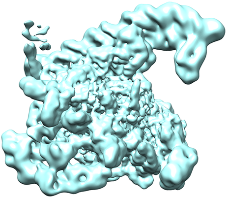 emd_12156.png
 http://ftp.pdbj.org/pub/emdb/structures/EMD-12156


 Z (Sec.)
 Y (Row.)
 X (Col.)


 Sample components
 Processing
 Electron microscopy
 FIELD EMISSION GUN

