 ムービー
ムービー コントローラー


+ データを開く

- 基本情報
基本情報
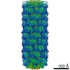
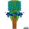
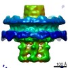
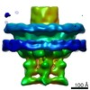
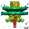
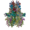
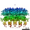
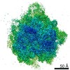
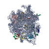
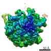
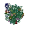
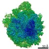
| 登録情報 | データベース: PDB / ID: 7b5i | ||||||
|---|---|---|---|---|---|---|---|
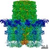
| タイトル | Cryo-EM structure of the contractile injection system cap complex from Anabaena PCC7120 | ||||||
 要素 要素 |
| ||||||
 キーワード キーワード | PROTEIN TRANSPORT / contractile tail / injection system / macromolecular machine / contractile protein | ||||||
| 機能・相同性 |  機能・相同性情報 機能・相同性情報 | ||||||
| 生物種 |  Nostoc sp. (バクテリア) Nostoc sp. (バクテリア) | ||||||
| 手法 | 電子顕微鏡法 / 単粒子再構成法 / クライオ電子顕微鏡法 / 解像度: 2.8 Å | ||||||
 データ登録者 データ登録者 | Eisenstein, F. / Weiss, G.L. / Pilhofer, M. | ||||||
 引用 引用 | ジャーナル: Nat Microbiol / 年: 2022 タイトル: Structure of a thylakoid-anchored contractile injection system in multicellular cyanobacteria. 著者: Gregor L Weiss / Fabian Eisenstein / Ann-Katrin Kieninger / Jingwei Xu / Hannah A Minas / Milena Gerber / Miki Feldmüller / Iris Maldener / Karl Forchhammer / Martin Pilhofer /    要旨: Contractile injection systems (CISs) mediate cell-cell interactions by phage tail-like structures, using two distinct modes of action: extracellular CISs are released into the medium, while type 6 ...Contractile injection systems (CISs) mediate cell-cell interactions by phage tail-like structures, using two distinct modes of action: extracellular CISs are released into the medium, while type 6 secretion systems (T6SSs) are attached to the cytoplasmic membrane and function upon cell-cell contact. Here, we characterized a CIS in the multicellular cyanobacterium Anabaena, with features distinct from extracellular CISs and T6SSs. Cryo-electron tomography of focused ion beam-milled cells revealed that CISs were anchored in thylakoid membrane stacks, facing the cell periphery. Single particle cryo-electron microscopy showed that this unique in situ localization was mediated by extensions of tail fibre and baseplate components. On stress, cyanobacteria induced the formation of ghost cells, presenting thylakoid-anchored CISs to the environment. Functional assays suggest that these CISs may mediate ghost cell formation and/or interactions of ghost cells with other organisms. Collectively, these data provide a framework for understanding the evolutionary re-engineering of CISs and potential roles of these CISs in cyanobacterial programmed cell death. | ||||||
| 履歴 |
|
- 構造の表示
構造の表示
| ムービー |
ムービービューア |
|---|---|
| 構造ビューア | 分子: MolmilJmol/JSmol |
- ダウンロードとリンク
-ダウンロード
| PDBx/mmCIF形式 | 7b5i.cif.gz | 1.3 MB | 表示 | PDBx/mmCIF形式 |
|---|---|---|---|---|
| PDB形式 | pdb7b5i.ent.gz | 表示 | PDB形式 | |
| PDBx/mmJSON形式 | 7b5i.json.gz | ツリー表示 | PDBx/mmJSON形式 | |
| その他 |  その他のダウンロード その他のダウンロード |
-検証レポート
| アーカイブディレクトリ | https://data.pdbj.org/pub/pdb/validation_reports/b5/7b5iftp://data.pdbj.org/pub/pdb/validation_reports/b5/7b5i | HTTPS FTP |
|---|
-関連構造データ


-リンク
 PDBj
PDBj- 集合体
集合体
| 登録構造単位 | 
|
|---|---|
| 1 |
|
-要素
| #1: タンパク質 | 分子量: 21935.064 Da / 分子数: 6 / 断片: cap protein Cis16A / 由来タイプ: 天然 / 詳細: cell culture 由来: (天然) Nostoc sp. (strain PCC 7120 / SAG 25.82 / UTEX 2576) (バクテリア)株: PCC 7120 / SAG 25.82 / UTEX 2576 / 参照: UniProt: Q8YRW5 #2: タンパク質 | 分子量: 45348.633 Da / 分子数: 6 / 断片: cap protein Cis16A / 由来タイプ: 天然 / 詳細: cell culture 由来: (天然) Nostoc sp. (strain PCC 7120 / SAG 25.82 / UTEX 2576) (バクテリア)株: PCC 7120 / SAG 25.82 / UTEX 2576 / 参照: UniProt: Q8YRW6 #3: タンパク質 | 分子量: 53236.160 Da / 分子数: 6 / 断片: cap protein Cis16A / 由来タイプ: 天然 / 詳細: cell culture 由来: (天然) Nostoc sp. (strain PCC 7120 / SAG 25.82 / UTEX 2576) (バクテリア)株: PCC 7120 / SAG 25.82 / UTEX 2576 / 参照: UniProt: Q8YRW7 #4: タンパク質 | 分子量: 16459.543 Da / 分子数: 12 / 断片: cap protein Cis16A / 由来タイプ: 天然 / 詳細: cell culture 由来: (天然) Nostoc sp. (strain PCC 7120 / SAG 25.82 / UTEX 2576) (バクテリア)株: PCC 7120 / SAG 25.82 / UTEX 2576 / 参照: UniProt: Q8YRW8 Has protein modification | Y | |
|---|
-実験情報
-実験
| 実験 | 手法: 電子顕微鏡法 |
|---|---|
| EM実験 | 試料の集合状態: PARTICLE / 3次元再構成法: 単粒子再構成法 |
- 試料調製
試料調製
| 構成要素 | 名称: Cap complex of the Anabaena PCC7120 contractile injection system タイプ: COMPLEX / Entity ID: all / 由来: NATURAL |
|---|---|
| 由来(天然) | 生物種: Nostoc sp. PCC 7120 = FACHB-418 (バクテリア) |
| 緩衝液 | pH: 7.5 |
| 試料 | 包埋: NO / シャドウイング: NO / 染色: NO / 凍結: YES |
| 試料支持 | グリッドの材料: COPPER / グリッドのタイプ: Quantifoil |
| 急速凍結 | 装置: FEI VITROBOT MARK IV / 凍結剤: ETHANE-PROPANE / 湿度: 100 % / 凍結前の試料温度: 281 K |
- 電子顕微鏡撮影
| 実験機器 |  モデル: Titan Krios / 画像提供: FEI Company |
|---|---|
| 顕微鏡 | モデル: FEI TITAN KRIOS |
| 電子銃 | 電子線源:  FIELD EMISSION GUN / 加速電圧: 300 kV / 照射モード: FLOOD BEAM FIELD EMISSION GUN / 加速電圧: 300 kV / 照射モード: FLOOD BEAM |
| 電子レンズ | モード: BRIGHT FIELD / Calibrated defocus min: 900 nm / 最大 デフォーカス(補正後): 1500 nm / Cs: 2.7 mm / C2レンズ絞り径: 50 µm |
| 試料ホルダ | 凍結剤: NITROGEN 試料ホルダーモデル: FEI TITAN KRIOS AUTOGRID HOLDER |
| 撮影 | 電子線照射量: 52 e/Å2 / 検出モード: COUNTING フィルム・検出器のモデル: GATAN K2 QUANTUM (4k x 4k) 撮影したグリッド数: 4 / 実像数: 19000 |
| 画像スキャン | 動画フレーム数/画像: 50 |
- 解析
解析
| EMソフトウェア |
| ||||||||||||||||||||||||||||||||
|---|---|---|---|---|---|---|---|---|---|---|---|---|---|---|---|---|---|---|---|---|---|---|---|---|---|---|---|---|---|---|---|---|---|
| CTF補正 | タイプ: PHASE FLIPPING AND AMPLITUDE CORRECTION | ||||||||||||||||||||||||||||||||
| 粒子像の選択 | 選択した粒子像数: 433028 | ||||||||||||||||||||||||||||||||
| 対称性 | 点対称性: C6 (6回回転対称) | ||||||||||||||||||||||||||||||||
| 3次元再構成 | 解像度: 2.8 Å / 解像度の算出法: FSC 0.143 CUT-OFF / 粒子像の数: 22598 / 対称性のタイプ: POINT |
