 Movie
Movie Controller

[English] 日本語
 Yorodumi
- PDB-7b4r: Structure of the 4'-phosphopantetheinyl transferase PptAb from My... -
+ Open data

- Basic information
| Entry | Database: PDB / ID: 7b4r | ||||||
|---|---|---|---|---|---|---|---|
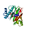
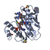
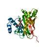
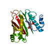
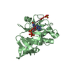
| Title | Structure of the 4'-phosphopantetheinyl transferase PptAb from Mycobacterium abscessus in complex with Coenzyme A and N-(2,6-diethylphenyl)-N'-(N-ethylcarbamimidoyl)urea | ||||||
 Components Components | Possible 4'-phosphopantetheinyl transferase | ||||||
 Keywords Keywords | TRANSFERASE / mycobacterium abscessus 4'-phosphopantetheinyl transferase complex / inhibition / amidino-urea | ||||||
| Function / homology |  Function and homology information Function and homology informationenterobactin synthetase complex / enterobactin biosynthetic process / holo-[acyl-carrier-protein] synthase activity / magnesium ion binding / plasma membrane Similarity search - Function | ||||||
| Biological species |  Mycobacteroides abscessus ATCC 19977 (bacteria) Mycobacteroides abscessus ATCC 19977 (bacteria) | ||||||
| Method |  X-RAY DIFFRACTION / SYNCHROTRON / FOURIER SYNTHESIS / Resolution: 1.4 Å X-RAY DIFFRACTION / SYNCHROTRON / FOURIER SYNTHESIS / Resolution: 1.4 Å | ||||||
 Authors Authors | Maveyraud, L. / Carivenc, C. / Blanger, C. / Mourey, L. | ||||||
| Funding support |  France, 1items France, 1items
| ||||||
 Citation Citation | Journal: Sci Rep / Year: 2021 Title: Phosphopantetheinyl transferase binding and inhibition by amidino-urea and hydroxypyrimidinethione compounds. Authors: Carivenc, C. / Maveyraud, L. / Blanger, C. / Ballereau, S. / Roy-Camille, C. / Nguyen, M.C. / Genisson, Y. / Guilhot, C. / Chalut, C. / Pedelacq, J.D. / Mourey, L. | ||||||
| History |
|
- Structure visualization
| Structure viewer | Molecule: MolmilJmol/JSmol |
|---|
- Downloads & links
-Download
| PDBx/mmCIF format | 7b4r.cif.gz | 163.8 KB | Display | PDBx/mmCIF format |
|---|---|---|---|---|
| PDB format | pdb7b4r.ent.gz | 118.5 KB | Display | PDB format |
| PDBx/mmJSON format | 7b4r.json.gz | Tree view | PDBx/mmJSON format | |
| Others |  Other downloads Other downloads |
-Validation report
| Arichive directory | https://data.pdbj.org/pub/pdb/validation_reports/b4/7b4rftp://data.pdbj.org/pub/pdb/validation_reports/b4/7b4r | HTTPS FTP |
|---|
-Related structure data
| Related structure data |  7b4sC  7bczC 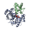 7bdwC 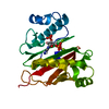 6qygS S: Starting model for refinement C: citing same article ( |
|---|---|
| Similar structure data |


-Links
 PDBj
PDBj


- Assembly
| Deposited unit | 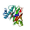
| ||||||||||||
|---|---|---|---|---|---|---|---|---|---|---|---|---|---|
| 1 |
| ||||||||||||
| Unit cell |
|
-Components
| #1: Protein | Mass: 25432.062 Da / Num. of mol.: 1 Source method: isolated from a genetically manipulated source Source: (gene. exp.) Mycobacteroides abscessus ATCC 19977 (bacteria)Gene: MAB_3117c / Production host: | ||||
|---|---|---|---|---|---|
| #2: Chemical | ChemComp-FD7 /   Mass: 262.351 Da / Num. of mol.: 1 / Source method: obtained synthetically / Formula: C14H22N4O / Feature type: SUBJECT OF INVESTIGATION Mass: 262.351 Da / Num. of mol.: 1 / Source method: obtained synthetically / Formula: C14H22N4O / Feature type: SUBJECT OF INVESTIGATION | ||||
| #3: Chemical | ChemComp-COA /   Mass: 767.534 Da / Num. of mol.: 1 / Source method: obtained synthetically / Formula: C21H36N7O16P3S Mass: 767.534 Da / Num. of mol.: 1 / Source method: obtained synthetically / Formula: C21H36N7O16P3S | ||||
| #4: Chemical |   Mass: 54.938 Da / Num. of mol.: 2 / Source method: obtained synthetically / Formula: Mn Mass: 54.938 Da / Num. of mol.: 2 / Source method: obtained synthetically / Formula: Mn#5: Water | ChemComp-HOH / |  Mass: 18.015 Da / Num. of mol.: 266 / Source method: isolated from a natural source / Formula: H2O Mass: 18.015 Da / Num. of mol.: 266 / Source method: isolated from a natural source / Formula: H2OHas ligand of interest | Y | |
-Experimental details
-Experiment
| Experiment | Method: X-RAY DIFFRACTION / Number of used crystals: 1 |
|---|
- Sample preparation
| Crystal | Density Matthews: 2.88 Å3/Da / Density % sol: 57.29 % |
|---|---|
| Crystal grow | Temperature: 285 K / Method: vapor diffusion, sitting drop / pH: 6.5 Details: 0.1 M LiSO4 0.1 M MES pH 6.5 20% (w/v) PEG 8K soaking 20h in 5 mM ligand 1% DMSO |
-Data collection
| Diffraction | Mean temperature: 100 K / Serial crystal experiment: N |
|---|---|
| Diffraction source | Source: SYNCHROTRON / Site: ALBA  / Beamline: XALOC / Wavelength: 0.97926 Å / Beamline: XALOC / Wavelength: 0.97926 Å |
| Detector | Type: DECTRIS PILATUS3 6M / Detector: PIXEL / Date: Mar 27, 2019 |
| Radiation | Protocol: SINGLE WAVELENGTH / Monochromatic (M) / Laue (L): M / Scattering type: x-ray |
| Radiation wavelength | Wavelength: 0.97926 Å / Relative weight: 1 |
| Reflection | Resolution: 1.4→46.44 Å / Num. obs: 57619 / % possible obs: 98.2 % / Redundancy: 6.4 % / Biso Wilson estimate: 22.82 Å2 / CC1/2: 0.999 / Rrim(I) all: 0.049 / Rsym value: 0.045 / Net I/σ(I): 15.8 |
| Reflection shell | Resolution: 1.4→1.48 Å / Redundancy: 6.6 % / Mean I/σ(I) obs: 1.3 / Num. unique obs: 9112 / CC1/2: 0.835 / Rrim(I) all: 1.18 / Rsym value: 1.087 / % possible all: 97.5 |
- Processing
| Software |
| ||||||||||||||||||||||||||||||||||||||||||||||||||||||||||||||||||||||||||||||||||||||||||||||||||||||||||||||||||||||||||||||||||||||||||||||||||||||||||
|---|---|---|---|---|---|---|---|---|---|---|---|---|---|---|---|---|---|---|---|---|---|---|---|---|---|---|---|---|---|---|---|---|---|---|---|---|---|---|---|---|---|---|---|---|---|---|---|---|---|---|---|---|---|---|---|---|---|---|---|---|---|---|---|---|---|---|---|---|---|---|---|---|---|---|---|---|---|---|---|---|---|---|---|---|---|---|---|---|---|---|---|---|---|---|---|---|---|---|---|---|---|---|---|---|---|---|---|---|---|---|---|---|---|---|---|---|---|---|---|---|---|---|---|---|---|---|---|---|---|---|---|---|---|---|---|---|---|---|---|---|---|---|---|---|---|---|---|---|---|---|---|---|---|---|---|
| Refinement | Method to determine structure: FOURIER SYNTHESIS Starting model: 6QYG Resolution: 1.4→46.44 Å / SU ML: 0.1733 / Cross valid method: FREE R-VALUE / σ(F): 1.36 / Phase error: 23.7179 Stereochemistry target values: GeoStd + Monomer Library + CDL v1.2
| ||||||||||||||||||||||||||||||||||||||||||||||||||||||||||||||||||||||||||||||||||||||||||||||||||||||||||||||||||||||||||||||||||||||||||||||||||||||||||
| Solvent computation | Shrinkage radii: 0.9 Å / VDW probe radii: 1.11 Å / Solvent model: FLAT BULK SOLVENT MODEL | ||||||||||||||||||||||||||||||||||||||||||||||||||||||||||||||||||||||||||||||||||||||||||||||||||||||||||||||||||||||||||||||||||||||||||||||||||||||||||
| Displacement parameters | Biso mean: 32.19 Å2 | ||||||||||||||||||||||||||||||||||||||||||||||||||||||||||||||||||||||||||||||||||||||||||||||||||||||||||||||||||||||||||||||||||||||||||||||||||||||||||
| Refinement step | Cycle: LAST / Resolution: 1.4→46.44 Å
| ||||||||||||||||||||||||||||||||||||||||||||||||||||||||||||||||||||||||||||||||||||||||||||||||||||||||||||||||||||||||||||||||||||||||||||||||||||||||||
| Refine LS restraints |
| ||||||||||||||||||||||||||||||||||||||||||||||||||||||||||||||||||||||||||||||||||||||||||||||||||||||||||||||||||||||||||||||||||||||||||||||||||||||||||
| LS refinement shell |
|
