Mass: 18.015 Da / Num. of mol.: 379 / Source method: isolated from a natural source / Formula: H2O
-
Experimental details
-
Experiment
Experiment
Method: X-RAY DIFFRACTION / Number of used crystals: 1
-
Sample preparation
Crystal
Density Matthews: 2.7 Å3/Da / Density % sol: 54.37 %
Crystal grow
Temperature: 296 K / Method: vapor diffusion, hanging drop / pH: 7 Details: 2.5 M NaCl, 100 mM Tris-HCl pH 7.0, 100 mM MgCl2, VAPOR DIFFUSION, HANGING DROP, temperature 296K
Redundancy: 2.9 % / Av σ(I) over netI: 27.88 / Number: 138717 / Rmerge(I) obs: 0.051 / Χ2: 1.01 / D res high: 2.3 Å / D res low: 100 Å / Num. obs: 47374 / % possible obs: 99.1
Diffraction reflection shell
Highest resolution (Å)
Lowest resolution (Å)
% possible obs (%)
ID
Rmerge(I) obs
Chi squared
Redundancy
4.96
100
98
1
0.028
1.019
2.9
3.93
4.96
99.5
1
0.03
1.036
3
3.44
3.93
99.7
1
0.038
0.98
3
3.12
3.44
99.6
1
0.05
1.007
3
2.9
3.12
99.6
1
0.072
1.035
3
2.73
2.9
99.5
1
0.096
1.05
3
2.59
2.73
99.2
1
0.136
1.018
3
2.48
2.59
99
1
0.187
0.979
2.9
2.38
2.48
98.6
1
0.224
0.964
2.9
2.3
2.38
98
1
0.264
0.981
2.8
Reflection
Resolution: 1.85→100 Å / Num. obs: 47833 / % possible obs: 99.7 % / Redundancy: 3.9 % / Rmerge(I) obs: 0.044 / Χ2: 1.023 / Net I/σ(I): 27.879
Reflection shell
Resolution (Å)
Redundancy (%)
Rmerge(I) obs
Num. unique all
Χ2
Diffraction-ID
% possible all
1.85-1.92
3.5
0.408
4687
1.186
1
99.6
1.92-1.99
3.9
0.292
4740
1.121
1
99.8
1.99-2.08
4
0.199
4700
1.062
1
99.8
2.08-2.19
4.1
0.123
4736
0.993
1
100
2.19-2.33
4
0.112
4732
1.017
1
99.7
2.33-2.51
4.1
0.067
4791
0.944
1
100
2.51-2.76
4.1
0.053
4768
0.947
1
100
2.76-3.16
4.1
0.037
4822
0.963
1
100
3.16-3.99
4
0.029
4843
1.045
1
99.8
3.99-100
3.8
0.021
5014
0.994
1
98.8
-
Phasing
Phasing
Method: MAD
Phasing set
ID
1
2
3
Phasing MAD
D res high: 2.36 Å / D res low: 30 Å / FOM : 0.47 / Reflection: 23362
Phasing MAD set
Clust-ID
Expt-ID
Set-ID
Wavelength (Å)
F double prime refined
F prime refined
1
3wavelength
1
0.9796
3.85
-8.32
1
3wavelength
2
1.02
0.54
-2.84
1
3wavelength
3
0.9797
0.5
-8.05
Phasing MAD set site
ID
Atom type symbol
B iso
Fract x
Fract y
Fract z
Occupancy
1
Se
14.631
0.801
0.24
0.216
0.629
2
Se
60
0.14
0.494
0.135
0.837
3
Se
60
0.16
0.582
0.186
0.761
4
Se
41.441
0.101
0.324
0.03
0.711
Phasing MAD shell
Resolution (Å)
FOM
Reflection
8.39-30
0.69
1192
5.33-8.39
0.66
1961
4.18-5.33
0.58
2503
3.54-4.18
0.54
2884
3.13-3.54
0.51
3266
2.84-3.13
0.45
3565
2.61-2.84
0.38
3843
2.43-2.61
0.28
4148
-
Processing
Software
Name
Version
Classification
NB
DENZO
datareduction
SCALEPACK
datascaling
SOLVE
2.1
phasing
RESOLVE
2.1
phasing
REFMAC
refmac_5.2.0005
refinement
PDB_EXTRACT
3.006
dataextraction
HKL-2000
datacollection
Refinement
Method to determine structure: MAD / Resolution: 1.85→59.98 Å / Cor.coef. Fo:Fc: 0.948 / Cor.coef. Fo:Fc free: 0.926 / WRfactor Rfree: 0.294 / WRfactor Rwork: 0.242 / Occupancy max: 1 / Occupancy min: 1 / SU B: 2.854 / SU ML: 0.088 / TLS residual ADP flag: LIKELY RESIDUAL / Cross valid method: THROUGHOUT / σ(F): 0 / ESU R: 0.138 / ESU R Free: 0.133 / Stereochemistry target values: MAXIMUM LIKELIHOOD / Details: HYDROGENS HAVE BEEN ADDED IN THE RIDING POSITIONS
Rfactor
Num. reflection
% reflection
Selection details
Rfree
0.243
2414
5.1 %
RANDOM
Rwork
0.206
-
-
-
obs
0.208
47768
99.55 %
-
Solvent computation
Ion probe radii: 0.8 Å / Shrinkage radii: 0.8 Å / VDW probe radii: 1.2 Å / Solvent model: MASK
In the structure databanks used in Yorodumi, some data are registered as the other names, "COVID-19 virus" and "2019-nCoV". Here are the details of the virus and the list of structure data.
Jan 31, 2019. EMDB accession codes are about to change! (news from PDBe EMDB page)
EMDB accession codes are about to change! (news from PDBe EMDB page)
The allocation of 4 digits for EMDB accession codes will soon come to an end. Whilst these codes will remain in use, new EMDB accession codes will include an additional digit and will expand incrementally as the available range of codes is exhausted. The current 4-digit format prefixed with “EMD-” (i.e. EMD-XXXX) will advance to a 5-digit format (i.e. EMD-XXXXX), and so on. It is currently estimated that the 4-digit codes will be depleted around Spring 2019, at which point the 5-digit format will come into force.
The EM Navigator/Yorodumi systems omit the EMD- prefix.
Related info.:Q: What is EMD? / ID/Accession-code notation in Yorodumi/EM Navigator
Yorodumi is a browser for structure data from EMDB, PDB, SASBDB, etc.
This page is also the successor to EM Navigator detail page, and also detail information page/front-end page for Omokage search.
The word "yorodu" (or yorozu) is an old Japanese word meaning "ten thousand". "mi" (miru) is to see.
Related info.:EMDB / PDB / SASBDB / Comparison of 3 databanks / Yorodumi Search / Aug 31, 2016. New EM Navigator & Yorodumi / Yorodumi Papers / Jmol/JSmol / Function and homology information / Changes in new EM Navigator and Yorodumi
 Movie
Movie Controller


 Open data
 Basic information
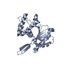
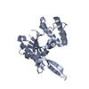
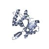
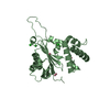
 Components
 Keywords
 Function and homology information
 Homo sapiens (human)
 X-RAY DIFFRACTION /
  Authors
 Citation
 Structure visualization
 Downloads & links
 Other downloads


 PDBj
PDBj


 Assembly


 Mass: 18.015 Da / Num. of mol.: 379 / Source method: isolated from a natural source / Formula: H2O
 Sample preparation
 / Beamline: 8.3.1 / Wavelength: 1.115872, 0.9796, 1.0200, 0.9797
 Processing