 Movie
Movie Controller

[English] 日本語
 Yorodumi
- PDB-7b4h: Structural basis of reactivation of oncogenic p53 mutants by a sm... -
+ Open data

- Basic information
| Entry | Database: PDB / ID: 7b4h | ||||||
|---|---|---|---|---|---|---|---|
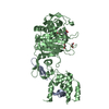
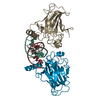
| Title | Structural basis of reactivation of oncogenic p53 mutants by a small molecule: methylene quinuclidinone (MQ). Human wild-type p53DBD bound to DNA and MQ: wt-DNA-MQ (III) | ||||||
 Components Components |
| ||||||
 Keywords Keywords | TRANSCRIPTION / P53 / TUMOR SUPPRESSOR / DNA BINDING PROTEIN / PROTEIN DNA COMPLEX / MICHAEL ACCEPTOR / MICHAEL REACTION / PROTEIN-DRUG COMPLEX / PROTEIN-DNA-DRUG COMPLEX / LOOP-SHEET-HELIX MOTIF / DNA TARGET / ACTIVATOR / HOOGSTEEN BASE-PAIRING | ||||||
| Function / homology |  Function and homology information Function and homology informationnegative regulation of helicase activity / Loss of function of TP53 in cancer due to loss of tetramerization ability / Regulation of TP53 Expression / signal transduction by p53 class mediator / negative regulation of G1 to G0 transition / negative regulation of glucose catabolic process to lactate via pyruvate / Transcriptional activation of cell cycle inhibitor p21 / regulation of intrinsic apoptotic signaling pathway by p53 class mediator / negative regulation of pentose-phosphate shunt / Activation of NOXA and translocation to mitochondria ...negative regulation of helicase activity / Loss of function of TP53 in cancer due to loss of tetramerization ability / Regulation of TP53 Expression / signal transduction by p53 class mediator / negative regulation of G1 to G0 transition / negative regulation of glucose catabolic process to lactate via pyruvate / Transcriptional activation of cell cycle inhibitor p21 / regulation of intrinsic apoptotic signaling pathway by p53 class mediator / negative regulation of pentose-phosphate shunt / Activation of NOXA and translocation to mitochondria / ATP-dependent DNA/DNA annealing activity / regulation of cell cycle G2/M phase transition / oligodendrocyte apoptotic process / negative regulation of miRNA processing / intrinsic apoptotic signaling pathway in response to hypoxia / positive regulation of thymocyte apoptotic process / oxidative stress-induced premature senescence / regulation of tissue remodeling / positive regulation of mitochondrial membrane permeability / germ cell nucleus / regulation of fibroblast apoptotic process / bone marrow development / circadian behavior / histone deacetylase regulator activity / positive regulation of programmed necrotic cell death / cellular response to actinomycin D / : / regulation of mitochondrial membrane permeability involved in apoptotic process / RUNX3 regulates CDKN1A transcription / T cell proliferation involved in immune response / TP53 Regulates Transcription of Death Receptors and Ligands / Activation of PUMA and translocation to mitochondria / TP53 regulates transcription of additional cell cycle genes whose exact role in the p53 pathway remain uncertain / mRNA transcription / negative regulation of glial cell proliferation / regulation of DNA damage response, signal transduction by p53 class mediator / Regulation of TP53 Activity through Association with Co-factors / negative regulation of neuroblast proliferation / Formation of Senescence-Associated Heterochromatin Foci (SAHF) / mitochondrial DNA repair / T cell lineage commitment / thymocyte apoptotic process / ER overload response / TP53 Regulates Transcription of Caspase Activators and Caspases / cardiac septum morphogenesis / B cell lineage commitment / entrainment of circadian clock by photoperiod / negative regulation of DNA replication / negative regulation of mitophagy / Zygotic genome activation (ZGA) / TP53 Regulates Transcription of Genes Involved in Cytochrome C Release / PI5P Regulates TP53 Acetylation / necroptotic process / negative regulation of telomere maintenance via telomerase / Association of TriC/CCT with target proteins during biosynthesis / positive regulation of release of cytochrome c from mitochondria / SUMOylation of transcription factors / TP53 regulates transcription of several additional cell death genes whose specific roles in p53-dependent apoptosis remain uncertain / rRNA transcription / negative regulation of reactive oxygen species metabolic process / TFIID-class transcription factor complex binding / Transcriptional Regulation by VENTX / intrinsic apoptotic signaling pathway by p53 class mediator / cellular response to UV-C / viral process / neuroblast proliferation / intrinsic apoptotic signaling pathway in response to endoplasmic reticulum stress / replicative senescence / positive regulation of RNA polymerase II transcription preinitiation complex assembly / Pyroptosis / intrinsic apoptotic signaling pathway in response to DNA damage by p53 class mediator / general transcription initiation factor binding / chromosome organization / positive regulation of execution phase of apoptosis / hematopoietic stem cell differentiation / type II interferon-mediated signaling pathway / embryonic organ development / response to X-ray / TP53 Regulates Transcription of Genes Involved in G1 Cell Cycle Arrest / somitogenesis / hematopoietic progenitor cell differentiation / negative regulation of stem cell proliferation / core promoter sequence-specific DNA binding / glial cell proliferation / negative regulation of fibroblast proliferation / cellular response to glucose starvation / mitophagy / cis-regulatory region sequence-specific DNA binding / Regulation of TP53 Activity through Acetylation / negative regulation of proteolysis / mitotic G1 DNA damage checkpoint signaling / positive regulation of intrinsic apoptotic signaling pathway / response to salt stress / cardiac muscle cell apoptotic process / transcription repressor complex / 14-3-3 protein binding / gastrulation / positive regulation of cardiac muscle cell apoptotic process / transforming growth factor beta receptor signaling pathway / MDM2/MDM4 family protein binding Similarity search - Function | ||||||
| Biological species |  Homo sapiens (human) Homo sapiens (human)synthetic construct (others) | ||||||
| Method |  X-RAY DIFFRACTION / SYNCHROTRON / MOLECULAR REPLACEMENT / molecular replacement / Resolution: 1.39 Å X-RAY DIFFRACTION / SYNCHROTRON / MOLECULAR REPLACEMENT / molecular replacement / Resolution: 1.39 Å | ||||||
 Authors Authors | Degtjarik, O. / Rozenberg, H. / Diskin-Posner, Y. / Shakked, Z. | ||||||
| Funding support | 1items
| ||||||
 Citation Citation | Journal: Nat Commun / Year: 2021 Title: Structural basis of reactivation of oncogenic p53 mutants by a small molecule: methylene quinuclidinone (MQ). Authors: Degtjarik, O. / Golovenko, D. / Diskin-Posner, Y. / Abrahmsen, L. / Rozenberg, H. / Shakked, Z. | ||||||
| History |
|
- Structure visualization
| Structure viewer | Molecule: MolmilJmol/JSmol |
|---|
- Downloads & links
-Download
| PDBx/mmCIF format | 7b4h.cif.gz | 131.8 KB | Display | PDBx/mmCIF format |
|---|---|---|---|---|
| PDB format | pdb7b4h.ent.gz | 80.9 KB | Display | PDB format |
| PDBx/mmJSON format | 7b4h.json.gz | Tree view | PDBx/mmJSON format | |
| Others |  Other downloads Other downloads |
-Validation report
| Arichive directory | https://data.pdbj.org/pub/pdb/validation_reports/b4/7b4hftp://data.pdbj.org/pub/pdb/validation_reports/b4/7b4h | HTTPS FTP |
|---|
-Related structure data
| Related structure data | 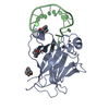 6zncC 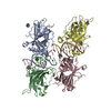 7b46C  7b47C  7b48C  7b49C  7b4aC  7b4bC  7b4cC  7b4dC 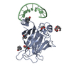 7b4eC 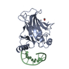 7b4fC 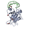 7b4gC 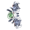 7b4nC 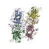 2ac0S C: citing same article ( S: Starting model for refinement |
|---|---|
| Similar structure data |


-Links
 PDBj
PDBj


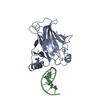
- Assembly
| Deposited unit | 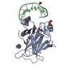
| ||||||||||||
|---|---|---|---|---|---|---|---|---|---|---|---|---|---|
| 1 | 
| ||||||||||||
| Unit cell |
|
-Components
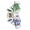
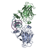
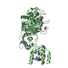
-Protein / DNA chain , 2 types, 2 molecules AB
| #1: Protein | Mass: 22505.582 Da / Num. of mol.: 1 / Fragment: p53 human DNA binding domain Source method: isolated from a genetically manipulated source Source: (gene. exp.) Homo sapiens (human) / Gene: TP53, P53 / Plasmid: pET-27-b / Production host:  |
|---|---|
| #2: DNA chain | Mass: 3664.380 Da / Num. of mol.: 1 / Fragment: p53 DNA target / Mutation: ' / Source method: obtained synthetically / Details: IDT: Integrated DNA Technology / Source: (synth.) synthetic construct (others) |
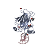
-Non-polymers , 7 types, 286 molecules 


| #3: Chemical | ChemComp-ZN /  Mass: 65.409 Da / Num. of mol.: 1 / Source method: obtained synthetically / Formula: Zn Mass: 65.409 Da / Num. of mol.: 1 / Source method: obtained synthetically / Formula: Zn | ||||||
|---|---|---|---|---|---|---|---|
| #4: Chemical | ChemComp-ACT /  Mass: 59.044 Da / Num. of mol.: 1 / Source method: obtained synthetically / Formula: C2H3O2 Mass: 59.044 Da / Num. of mol.: 1 / Source method: obtained synthetically / Formula: C2H3O2 | ||||||
| #5: Chemical | ChemComp-EDO /  Mass: 62.068 Da / Num. of mol.: 1 / Source method: obtained synthetically / Formula: C2H6O2 Mass: 62.068 Da / Num. of mol.: 1 / Source method: obtained synthetically / Formula: C2H6O2 | ||||||
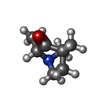
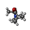
| #6: Chemical |  Mass: 139.195 Da / Num. of mol.: 2 / Source method: obtained synthetically / Formula: C8H13NO / Feature type: SUBJECT OF INVESTIGATION Mass: 139.195 Da / Num. of mol.: 2 / Source method: obtained synthetically / Formula: C8H13NO / Feature type: SUBJECT OF INVESTIGATION#7: Chemical | ChemComp-QNN / ( |  Mass: 139.195 Da / Num. of mol.: 1 / Source method: obtained synthetically / Formula: C8H13NO / Feature type: SUBJECT OF INVESTIGATION Mass: 139.195 Da / Num. of mol.: 1 / Source method: obtained synthetically / Formula: C8H13NO / Feature type: SUBJECT OF INVESTIGATION#8: Chemical | ChemComp-FMT /  Mass: 46.025 Da / Num. of mol.: 4 / Source method: obtained synthetically / Formula: CH2O2 Mass: 46.025 Da / Num. of mol.: 4 / Source method: obtained synthetically / Formula: CH2O2#9: Water | ChemComp-HOH / | Mass: 18.015 Da / Num. of mol.: 276 / Source method: isolated from a natural source / Formula: H2O |
-Details
| Has ligand of interest | Y |
|---|---|
| Has protein modification | Y |
| Nonpolymer details | Two enantiomers, QNN and QN8 are produced by addition reaction of the michael acceptor compound "2- ...Two enantiomers, QNN and QN8 are produced by addition reaction of the michael acceptor compound "2-methylenequinuclidin-3-one" with cysteine or lysine. As such, QNN and QN8 bind covalently to the thiol group of cysteine or amino group of lysine. The chiral definitions of QNN and QN8 bound to cysteines are reversed to that of the pseudo free ligands. |
-Experimental details
-Experiment
| Experiment | Method: X-RAY DIFFRACTION / Number of used crystals: 1 |
|---|
- Sample preparation
| Crystal | Density Matthews: 2.24 Å3/Da / Density % sol: 45.14 % / Mosaicity: 0.374 ° |
|---|---|
| Crystal grow | Temperature: 292 K / Method: evaporation Details: Protein/DNA ratio 1:2.4, 0.1M TRIS pH=8.5, 2% Tacsimate(TM) pH=8.0, 16% w/v PEG 3350 |
-Data collection
| Diffraction | Mean temperature: 100 K / Serial crystal experiment: N | |||||||||||||||||||||||||||||||||||||||||||||||||||||||||||||||||||||||||||||||||||||||||||||||||||||||||||||||||||||||||||||||||||||||||||||||||||||||||||||||||||||||||||||||||||||||||||||
|---|---|---|---|---|---|---|---|---|---|---|---|---|---|---|---|---|---|---|---|---|---|---|---|---|---|---|---|---|---|---|---|---|---|---|---|---|---|---|---|---|---|---|---|---|---|---|---|---|---|---|---|---|---|---|---|---|---|---|---|---|---|---|---|---|---|---|---|---|---|---|---|---|---|---|---|---|---|---|---|---|---|---|---|---|---|---|---|---|---|---|---|---|---|---|---|---|---|---|---|---|---|---|---|---|---|---|---|---|---|---|---|---|---|---|---|---|---|---|---|---|---|---|---|---|---|---|---|---|---|---|---|---|---|---|---|---|---|---|---|---|---|---|---|---|---|---|---|---|---|---|---|---|---|---|---|---|---|---|---|---|---|---|---|---|---|---|---|---|---|---|---|---|---|---|---|---|---|---|---|---|---|---|---|---|---|---|---|---|---|---|
| Diffraction source | Source: SYNCHROTRON / Site: ESRF  / Beamline: ID23-1 / Wavelength: 0.976 Å / Beamline: ID23-1 / Wavelength: 0.976 Å | |||||||||||||||||||||||||||||||||||||||||||||||||||||||||||||||||||||||||||||||||||||||||||||||||||||||||||||||||||||||||||||||||||||||||||||||||||||||||||||||||||||||||||||||||||||||||||||
| Detector | Type: DECTRIS PILATUS 6M / Detector: PIXEL / Date: Apr 27, 2014 | |||||||||||||||||||||||||||||||||||||||||||||||||||||||||||||||||||||||||||||||||||||||||||||||||||||||||||||||||||||||||||||||||||||||||||||||||||||||||||||||||||||||||||||||||||||||||||||
| Radiation | Protocol: SINGLE WAVELENGTH / Monochromatic (M) / Laue (L): M / Scattering type: x-ray | |||||||||||||||||||||||||||||||||||||||||||||||||||||||||||||||||||||||||||||||||||||||||||||||||||||||||||||||||||||||||||||||||||||||||||||||||||||||||||||||||||||||||||||||||||||||||||||
| Radiation wavelength | Wavelength: 0.976 Å / Relative weight: 1 | |||||||||||||||||||||||||||||||||||||||||||||||||||||||||||||||||||||||||||||||||||||||||||||||||||||||||||||||||||||||||||||||||||||||||||||||||||||||||||||||||||||||||||||||||||||||||||||
| Reflection | Resolution: 1.39→49 Å / Num. obs: 46808 / % possible obs: 99.7 % / Redundancy: 7.3 % / Rmerge(I) obs: 0.093 / Rpim(I) all: 0.036 / Rrim(I) all: 0.1 / Χ2: 1.011 / Net I/σ(I): 6.1 / Num. measured all: 339531 | |||||||||||||||||||||||||||||||||||||||||||||||||||||||||||||||||||||||||||||||||||||||||||||||||||||||||||||||||||||||||||||||||||||||||||||||||||||||||||||||||||||||||||||||||||||||||||||
| Reflection shell | Diffraction-ID: 1
|
-Phasing
| Phasing | Method: molecular replacement |
|---|
- Processing
| Software |
| ||||||||||||||||||||||||||||||||||||||||||||||||||||||||||||||||||||||||||||||||||||||||||||||||||||||||||||||||||||||||||||||
|---|---|---|---|---|---|---|---|---|---|---|---|---|---|---|---|---|---|---|---|---|---|---|---|---|---|---|---|---|---|---|---|---|---|---|---|---|---|---|---|---|---|---|---|---|---|---|---|---|---|---|---|---|---|---|---|---|---|---|---|---|---|---|---|---|---|---|---|---|---|---|---|---|---|---|---|---|---|---|---|---|---|---|---|---|---|---|---|---|---|---|---|---|---|---|---|---|---|---|---|---|---|---|---|---|---|---|---|---|---|---|---|---|---|---|---|---|---|---|---|---|---|---|---|---|---|---|---|
| Refinement | Method to determine structure: MOLECULAR REPLACEMENT Starting model: 2AC0 Resolution: 1.39→47.07 Å / SU ML: 0.1641 / Cross valid method: FREE R-VALUE / σ(F): 1.36 / Phase error: 19.6678 Stereochemistry target values: GeoStd + Monomer Library + CDL v1.2
| ||||||||||||||||||||||||||||||||||||||||||||||||||||||||||||||||||||||||||||||||||||||||||||||||||||||||||||||||||||||||||||||
| Solvent computation | Shrinkage radii: 0.9 Å / VDW probe radii: 1.11 Å / Solvent model: FLAT BULK SOLVENT MODEL | ||||||||||||||||||||||||||||||||||||||||||||||||||||||||||||||||||||||||||||||||||||||||||||||||||||||||||||||||||||||||||||||
| Displacement parameters | Biso mean: 24.84 Å2 | ||||||||||||||||||||||||||||||||||||||||||||||||||||||||||||||||||||||||||||||||||||||||||||||||||||||||||||||||||||||||||||||
| Refinement step | Cycle: LAST / Resolution: 1.39→47.07 Å
| ||||||||||||||||||||||||||||||||||||||||||||||||||||||||||||||||||||||||||||||||||||||||||||||||||||||||||||||||||||||||||||||
| Refine LS restraints |
| ||||||||||||||||||||||||||||||||||||||||||||||||||||||||||||||||||||||||||||||||||||||||||||||||||||||||||||||||||||||||||||||
| LS refinement shell |
|
