 Movie
Movie Controller


+ Open data

- Basic information
| Entry | Database: PDB / ID: 7b30 | ||||||
|---|---|---|---|---|---|---|---|
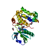
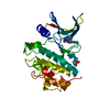
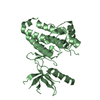
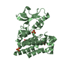
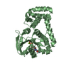
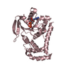
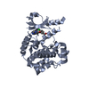
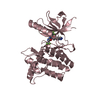
| Title | MST3 in complex with compound G-5555 | ||||||
 Components Components | Serine/threonine-protein kinase 24 | ||||||
 Keywords Keywords | TRANSFERASE / kinase inhibitors / structure-based drug design / SIK2 inhibitor / Structural Genomics Consortium / SGC | ||||||
| Function / homology |  Function and homology information Function and homology informationregulation of axon regeneration / Apoptotic execution phase / FAR/SIN/STRIPAK complex / intrinsic apoptotic signaling pathway in response to oxidative stress / execution phase of apoptosis / Apoptotic cleavage of cellular proteins / negative regulation of cell migration / cellular response to starvation / protein autophosphorylation / cellular response to oxidative stress ...regulation of axon regeneration / Apoptotic execution phase / FAR/SIN/STRIPAK complex / intrinsic apoptotic signaling pathway in response to oxidative stress / execution phase of apoptosis / Apoptotic cleavage of cellular proteins / negative regulation of cell migration / cellular response to starvation / protein autophosphorylation / cellular response to oxidative stress / protein phosphorylation / protein kinase activity / non-specific serine/threonine protein kinase / intracellular signal transduction / cadherin binding / protein serine kinase activity / protein serine/threonine kinase activity / nucleolus / Golgi apparatus / signal transduction / extracellular exosome / nucleoplasm / ATP binding / membrane / metal ion binding / nucleus / cytosol / cytoplasm Similarity search - Function | ||||||
| Biological species |  Homo sapiens (human) Homo sapiens (human) | ||||||
| Method |  X-RAY DIFFRACTION / SYNCHROTRON / MOLECULAR REPLACEMENT / Resolution: 2.1 Å X-RAY DIFFRACTION / SYNCHROTRON / MOLECULAR REPLACEMENT / Resolution: 2.1 Å | ||||||
 Authors Authors | Tesch, R. / Rak, M. / Joerger, A.C. / Knapp, S. / Structural Genomics Consortium (SGC) | ||||||
| Funding support |  Germany, 1items Germany, 1items
| ||||||
 Citation Citation | Journal: J.Med.Chem. / Year: 2021 Title: Structure-Based Design of Selective Salt-Inducible Kinase Inhibitors. Authors: Tesch, R. / Rak, M. / Raab, M. / Berger, L.M. / Kronenberger, T. / Joerger, A.C. / Berger, B.T. / Abdi, I. / Hanke, T. / Poso, A. / Strebhardt, K. / Sanhaji, M. / Knapp, S. | ||||||
| History |
|
- Structure visualization
| Structure viewer | Molecule: MolmilJmol/JSmol |
|---|
- Downloads & links
-Download
| PDBx/mmCIF format | 7b30.cif.gz | 143.1 KB | Display | PDBx/mmCIF format |
|---|---|---|---|---|
| PDB format | pdb7b30.ent.gz | 92.6 KB | Display | PDB format |
| PDBx/mmJSON format | 7b30.json.gz | Tree view | PDBx/mmJSON format | |
| Others |  Other downloads Other downloads |
-Validation report
| Arichive directory | https://data.pdbj.org/pub/pdb/validation_reports/b3/7b30ftp://data.pdbj.org/pub/pdb/validation_reports/b3/7b30 | HTTPS FTP |
|---|
-Related structure data
| Related structure data |  7b31C  7b32C  7b33C  7b34C  7b35C  7b36C  3zhpS S: Starting model for refinement C: citing same article ( |
|---|---|
| Similar structure data |


-Links
 PDBj
PDBj
- Assembly
| Deposited unit | 
| ||||||||||||
|---|---|---|---|---|---|---|---|---|---|---|---|---|---|
| 1 |
| ||||||||||||
| Unit cell |
|
-Components
| #1: Protein | Mass: 34559.648 Da / Num. of mol.: 1 Source method: isolated from a genetically manipulated source Source: (gene. exp.) Homo sapiens (human) / Gene: STK24, MST3, STK3 / Production host:  References: UniProt: Q9Y6E0, non-specific serine/threonine protein kinase |
|---|---|
| #2: Chemical | ChemComp-59T /   Mass: 492.957 Da / Num. of mol.: 1 / Source method: obtained synthetically / Formula: C25H25ClN6O3 / Feature type: SUBJECT OF INVESTIGATION Mass: 492.957 Da / Num. of mol.: 1 / Source method: obtained synthetically / Formula: C25H25ClN6O3 / Feature type: SUBJECT OF INVESTIGATION |
| #3: Water | ChemComp-HOH /  Mass: 18.015 Da / Num. of mol.: 108 / Source method: isolated from a natural source / Formula: H2O Mass: 18.015 Da / Num. of mol.: 108 / Source method: isolated from a natural source / Formula: H2O |
| Has ligand of interest | Y |
-Experimental details
-Experiment
| Experiment | Method: X-RAY DIFFRACTION / Number of used crystals: 1 |
|---|
- Sample preparation
| Crystal | Density Matthews: 2.6 Å3/Da / Density % sol: 52.74 % |
|---|---|
| Crystal grow | Temperature: 293 K / Method: vapor diffusion, sitting drop Details: Protein solution: 10 mg/ml in buffer 25 mM HEPES pH 7.5, 200 mM NaCl, 5% glycerol and 0.5 mM TCEP. Reservoir: 10% PEG 6000, 0.1M HEPES pH 7.0. |
-Data collection
| Diffraction | Mean temperature: 100 K / Serial crystal experiment: N |
|---|---|
| Diffraction source | Source: SYNCHROTRON / Site: SLS  / Beamline: X06SA / Wavelength: 1.00004 Å / Beamline: X06SA / Wavelength: 1.00004 Å |
| Detector | Type: DECTRIS EIGER X 16M / Detector: PIXEL / Date: Mar 7, 2020 |
| Radiation | Protocol: SINGLE WAVELENGTH / Monochromatic (M) / Laue (L): M / Scattering type: x-ray |
| Radiation wavelength | Wavelength: 1.00004 Å / Relative weight: 1 |
| Reflection | Resolution: 2.1→39.64 Å / Num. obs: 20236 / % possible obs: 97 % / Redundancy: 6 % / Biso Wilson estimate: 38.68 Å2 / CC1/2: 0.997 / Rpim(I) all: 0.03 / Net I/σ(I): 12.6 |
| Reflection shell | Resolution: 2.1→2.16 Å / Redundancy: 4.7 % / Mean I/σ(I) obs: 3.6 / Num. unique obs: 1341 / CC1/2: 0.881 / Rpim(I) all: 0.19 / % possible all: 79.3 |
- Processing
| Software |
| |||||||||||||||||||||||||||||||||||||||||||||||||||||||||||||||||||||||||||
|---|---|---|---|---|---|---|---|---|---|---|---|---|---|---|---|---|---|---|---|---|---|---|---|---|---|---|---|---|---|---|---|---|---|---|---|---|---|---|---|---|---|---|---|---|---|---|---|---|---|---|---|---|---|---|---|---|---|---|---|---|---|---|---|---|---|---|---|---|---|---|---|---|---|---|---|---|
| Refinement | Method to determine structure: MOLECULAR REPLACEMENT Starting model: 3ZHP Resolution: 2.1→39.64 Å / SU ML: 0.1967 / Cross valid method: FREE R-VALUE / σ(F): 1.34 / Phase error: 20.5467 Stereochemistry target values: GeoStd + Monomer Library + CDL v1.2
| |||||||||||||||||||||||||||||||||||||||||||||||||||||||||||||||||||||||||||
| Solvent computation | Shrinkage radii: 0.9 Å / VDW probe radii: 1.11 Å / Solvent model: FLAT BULK SOLVENT MODEL | |||||||||||||||||||||||||||||||||||||||||||||||||||||||||||||||||||||||||||
| Displacement parameters | Biso mean: 45.91 Å2 | |||||||||||||||||||||||||||||||||||||||||||||||||||||||||||||||||||||||||||
| Refinement step | Cycle: LAST / Resolution: 2.1→39.64 Å
| |||||||||||||||||||||||||||||||||||||||||||||||||||||||||||||||||||||||||||
| Refine LS restraints |
| |||||||||||||||||||||||||||||||||||||||||||||||||||||||||||||||||||||||||||
| LS refinement shell |
| |||||||||||||||||||||||||||||||||||||||||||||||||||||||||||||||||||||||||||
| Refinement TLS params. | Method: refined / Refine-ID: X-RAY DIFFRACTION
| |||||||||||||||||||||||||||||||||||||||||||||||||||||||||||||||||||||||||||
| Refinement TLS group | Refine-ID: X-RAY DIFFRACTION / Auth asym-ID: A / Label asym-ID: A
|
