| 登録情報 | データベース: PDB / ID: 7b1h
|
|---|
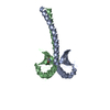
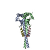
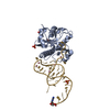
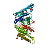
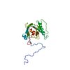
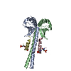
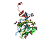
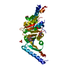
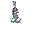
| タイトル | Monoclinic P21 Structure of Human Mad1 C-terminal Domain in Complex with Phosphorylated Bub1 CD1 Domain |
|---|
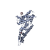
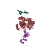
 要素 要素 | - Mitotic checkpoint serine/threonine-protein kinase BUB1
- Mitotic spindle assembly checkpoint protein MAD1
|
|---|
 キーワード キーワード | CELL CYCLE / Mad1 / Bub1 / spindle assembly checkpoint / mitotic checkpoint complex |
|---|
| 機能・相同性 |  機能・相同性情報 機能・相同性情報
MAD1 complex / positive regulation of maintenance of mitotic sister chromatid cohesion, centromeric / histone H2A kinase activity / deactivation of mitotic spindle assembly checkpoint / mitotic spindle assembly checkpoint MAD1-MAD2 complex / regulation of sister chromatid cohesion / regulation of chromosome segregation / positive regulation of mitotic cell cycle spindle assembly checkpoint / meiotic sister chromatid cohesion, centromeric / kinetochore binding ...MAD1 complex / positive regulation of maintenance of mitotic sister chromatid cohesion, centromeric / histone H2A kinase activity / deactivation of mitotic spindle assembly checkpoint / mitotic spindle assembly checkpoint MAD1-MAD2 complex / regulation of sister chromatid cohesion / regulation of chromosome segregation / positive regulation of mitotic cell cycle spindle assembly checkpoint / meiotic sister chromatid cohesion, centromeric / kinetochore binding / nuclear pore nuclear basket / outer kinetochore / regulation of metaphase plate congression / attachment of mitotic spindle microtubules to kinetochore / mitotic spindle assembly checkpoint signaling / negative regulation of T cell proliferation / Amplification of signal from unattached kinetochores via a MAD2 inhibitory signal / Mitotic Prometaphase / EML4 and NUDC in mitotic spindle formation / Resolution of Sister Chromatid Cohesion / thymus development / chromosome segregation / RHO GTPases Activate Formins / kinetochore / spindle / spindle pole / mitotic spindle / Separation of Sister Chromatids / nuclear envelope / non-specific serine/threonine protein kinase / protein kinase activity / cell division / protein serine kinase activity / intracellular membrane-bounded organelle / protein serine/threonine kinase activity / apoptotic process / centrosome / nucleoplasm / ATP binding / identical protein binding / nucleus / membrane / cytoplasm / cytosol類似検索 - 分子機能 Spindle assembly checkpoint component Mad1 / Mitotic checkpoint protein / Mad3/Bub1 homology region 1 / Mitotic spindle checkpoint protein Bub1/Mad3 / Mad3/BUB1 homology region 1 / BUB1 N-terminal domain profile. / Mad3/BUB1 hoMad3/BUB1 homology region 1 / Serine/threonine-protein kinase, active site / Serine/Threonine protein kinases active-site signature. / Protein kinase domain ...Spindle assembly checkpoint component Mad1 / Mitotic checkpoint protein / Mad3/Bub1 homology region 1 / Mitotic spindle checkpoint protein Bub1/Mad3 / Mad3/BUB1 homology region 1 / BUB1 N-terminal domain profile. / Mad3/BUB1 hoMad3/BUB1 homology region 1 / Serine/threonine-protein kinase, active site / Serine/Threonine protein kinases active-site signature. / Protein kinase domain / Serine/Threonine protein kinases, catalytic domain / Protein kinase, ATP binding site / Protein kinases ATP-binding region signature. / Protein kinase domain profile. / Protein kinase domain / Protein kinase-like domain superfamily類似検索 - ドメイン・相同性 Mitotic checkpoint serine/threonine-protein kinase BUB1 / Mitotic spindle assembly checkpoint protein MAD1類似検索 - 構成要素 |
|---|
| 生物種 |  Homo sapiens (ヒト) Homo sapiens (ヒト) |
|---|
| 手法 |  X線回折 / シンクロトロン / 分子置換 / 解像度: 2.4 Å X線回折 / シンクロトロン / 分子置換 / 解像度: 2.4 Å |
|---|
 データ登録者 データ登録者 | Fischer, E. / Bellini, D. / Barford, D. |
|---|
| 資金援助 |  英国, 2件 英国, 2件 | 組織 | 認可番号 | 国 |
|---|
| Medical Research Council (MRC, United Kingdom) | MC_UP_1201/6 | 英国 | | Cancer Research UK | C576/A14109 | 英国 |
|
|---|
 引用 引用 | ジャーナル: Embo Rep. / 年: 2021
タイトル: Molecular mechanism of Mad1 kinetochore targeting by phosphorylated Bub1.
著者: Fischer, E.S. / Yu, C.W.H. / Bellini, D. / McLaughlin, S.H. / Orr, C.M. / Wagner, A. / Freund, S.M.V. / Barford, D. |
|---|
| 履歴 | | 登録 | 2020年11月24日 | 登録サイト: PDBE / 処理サイト: PDBE |
|---|
| 改定 1.0 | 2021年3月17日 | Provider: repository / タイプ: Initial release |
|---|
| 改定 1.1 | 2021年6月2日 | Group: Database references / カテゴリ: citation / citation_author
Item: _citation.country / _citation.journal_abbrev ..._citation.country / _citation.journal_abbrev / _citation.journal_id_CSD / _citation.journal_id_ISSN / _citation.page_first / _citation.page_last / _citation.pdbx_database_id_DOI / _citation.pdbx_database_id_PubMed / _citation.title / _citation.year / _citation_author.identifier_ORCID / _citation_author.name |
|---|
| 改定 1.2 | 2021年7月14日 | Group: Database references / カテゴリ: citation / Item: _citation.journal_volume |
|---|
| 改定 1.3 | 2024年1月31日 | Group: Data collection / Database references / Refinement description
カテゴリ: chem_comp_atom / chem_comp_bond ...chem_comp_atom / chem_comp_bond / database_2 / pdbx_initial_refinement_model
Item: _database_2.pdbx_DOI / _database_2.pdbx_database_accession |
|---|
| 改定 1.4 | 2024年11月13日 | Group: Structure summary
カテゴリ: pdbx_entry_details / pdbx_modification_feature
Item: _pdbx_entry_details.has_protein_modification |
|---|
|
|---|
 ムービー
ムービー コントローラー


 データを開く
 基本情報
基本情報 構造の表示
構造の表示 ダウンロードとリンク
 その他のダウンロード


 PDBj
PDBj


 集合体
集合体


 分子量: 18.015 Da / 分子数: 12 / 由来タイプ: 天然 / 式: H2O
 試料調製
試料調製 解析
解析