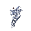
 Movie
Movie Controller

[English] 日本語
 Yorodumi
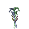
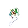
- PDB-7b1h: Monoclinic P21 Structure of Human Mad1 C-terminal Domain in Compl... -
+ Open data

- Basic information
| Entry | Database: PDB / ID: 7b1h | |||||||||
|---|---|---|---|---|---|---|---|---|---|---|
| Title | Monoclinic P21 Structure of Human Mad1 C-terminal Domain in Complex with Phosphorylated Bub1 CD1 Domain | |||||||||
 Components Components |
| |||||||||
 Keywords Keywords | CELL CYCLE / Mad1 / Bub1 / spindle assembly checkpoint / mitotic checkpoint complex | |||||||||
| Function / homology |  Function and homology information Function and homology informationMAD1 complex / positive regulation of maintenance of mitotic sister chromatid cohesion, centromeric / histone H2A kinase activity / deactivation of mitotic spindle assembly checkpoint / mitotic spindle assembly checkpoint MAD1-MAD2 complex / regulation of sister chromatid cohesion / regulation of chromosome segregation / meiotic sister chromatid cohesion, centromeric / positive regulation of mitotic cell cycle spindle assembly checkpoint / kinetochore binding ...MAD1 complex / positive regulation of maintenance of mitotic sister chromatid cohesion, centromeric / histone H2A kinase activity / deactivation of mitotic spindle assembly checkpoint / mitotic spindle assembly checkpoint MAD1-MAD2 complex / regulation of sister chromatid cohesion / regulation of chromosome segregation / meiotic sister chromatid cohesion, centromeric / positive regulation of mitotic cell cycle spindle assembly checkpoint / kinetochore binding / nuclear pore nuclear basket / outer kinetochore / regulation of metaphase plate congression / attachment of mitotic spindle microtubules to kinetochore / mitotic spindle assembly checkpoint signaling / negative regulation of T cell proliferation / Amplification of signal from unattached kinetochores via a MAD2 inhibitory signal / Mitotic Prometaphase / EML4 and NUDC in mitotic spindle formation / thymus development / Resolution of Sister Chromatid Cohesion / chromosome segregation / RHO GTPases Activate Formins / kinetochore / spindle / spindle pole / mitotic spindle / Separation of Sister Chromatids / nuclear envelope / protein kinase activity / non-specific serine/threonine protein kinase / protein serine kinase activity / cell division / protein serine/threonine kinase activity / apoptotic process / centrosome / nucleoplasm / ATP binding / membrane / identical protein binding / nucleus / cytoplasm / cytosol Similarity search - Function | |||||||||
| Biological species |  Homo sapiens (human) Homo sapiens (human) | |||||||||
| Method |  X-RAY DIFFRACTION / SYNCHROTRON / MOLECULAR REPLACEMENT / Resolution: 2.4 Å X-RAY DIFFRACTION / SYNCHROTRON / MOLECULAR REPLACEMENT / Resolution: 2.4 Å | |||||||||
 Authors Authors | Fischer, E. / Bellini, D. / Barford, D. | |||||||||
| Funding support |  United Kingdom, 2items United Kingdom, 2items
| |||||||||
 Citation Citation | Journal: Embo Rep. / Year: 2021 Title: Molecular mechanism of Mad1 kinetochore targeting by phosphorylated Bub1. Authors: Fischer, E.S. / Yu, C.W.H. / Bellini, D. / McLaughlin, S.H. / Orr, C.M. / Wagner, A. / Freund, S.M.V. / Barford, D. | |||||||||
| History |
|
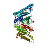
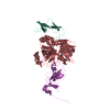
- Structure visualization
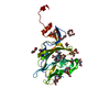
| Structure viewer | Molecule: MolmilJmol/JSmol |
|---|
- Downloads & links
-Download
| PDBx/mmCIF format | 7b1h.cif.gz | 124.4 KB | Display | PDBx/mmCIF format |
|---|---|---|---|---|
| PDB format | pdb7b1h.ent.gz | 96.9 KB | Display | PDB format |
| PDBx/mmJSON format | 7b1h.json.gz | Tree view | PDBx/mmJSON format | |
| Others |  Other downloads Other downloads |
-Validation report
| Arichive directory | https://data.pdbj.org/pub/pdb/validation_reports/b1/7b1hftp://data.pdbj.org/pub/pdb/validation_reports/b1/7b1h | HTTPS FTP |
|---|
-Related structure data
| Related structure data |  7b1fC  7b1jC 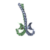 4dzoS S: Starting model for refinement C: citing same article ( |
|---|---|
| Similar structure data |

-Links
 PDBj
PDBj


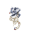
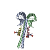
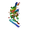
- Assembly
| Deposited unit | 
| ||||||||
|---|---|---|---|---|---|---|---|---|---|
| 1 | 
| ||||||||
| 2 | 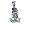
| ||||||||
| Unit cell |
|
-Components
| #1: Protein | Mass: 13961.832 Da / Num. of mol.: 4 Source method: isolated from a genetically manipulated source Source: (gene. exp.) Homo sapiens (human) / Gene: MAD1L1, MAD1, TXBP181 / Production host:  #2: Protein/peptide | Mass: 3023.290 Da / Num. of mol.: 4 / Source method: obtained synthetically / Source: (synth.) Homo sapiens (human)References: UniProt: O43683, non-specific serine/threonine protein kinase #3: Water | ChemComp-HOH / |  Mass: 18.015 Da / Num. of mol.: 12 / Source method: isolated from a natural source / Formula: H2O Mass: 18.015 Da / Num. of mol.: 12 / Source method: isolated from a natural source / Formula: H2OHas ligand of interest | Y | Has protein modification | Y | |
|---|
-Experimental details
-Experiment
| Experiment | Method: X-RAY DIFFRACTION / Number of used crystals: 1 |
|---|
- Sample preparation
| Crystal | Density Matthews: 2.79 Å3/Da / Density % sol: 55.85 % |
|---|---|
| Crystal grow | Temperature: 293 K / Method: vapor diffusion, sitting drop / pH: 7.5 Details: 8% Isopropanol, 20% PEG 4000, 0.1 M Na HEPES, pH 7.5 |
-Data collection
| Diffraction | Mean temperature: 100 K / Serial crystal experiment: N |
|---|---|
| Diffraction source | Source: SYNCHROTRON / Site: Diamond / Beamline: I04 / Wavelength: 0.9795 Å |
| Detector | Type: DECTRIS EIGER X 16M / Detector: PIXEL / Date: Jul 21, 2020 |
| Radiation | Protocol: SINGLE WAVELENGTH / Monochromatic (M) / Laue (L): M / Scattering type: x-ray |
| Radiation wavelength | Wavelength: 0.9795 Å / Relative weight: 1 |
| Reflection | Resolution: 2.4→39.43 Å / Num. obs: 100346 / % possible obs: 99.56 % / Redundancy: 3.5 % / CC1/2: 1 / Rmerge(I) obs: 0.06 / Rpim(I) all: 0.04 / Rrim(I) all: 0.07 / Net I/σ(I): 14.5 |
| Reflection shell | Resolution: 2.4→2.486 Å / Redundancy: 3.4 % / Rmerge(I) obs: 0.06 / Mean I/σ(I) obs: 1.7 / Num. unique obs: 4893 / CC1/2: 0.68 / Rpim(I) all: 0.04 / Rrim(I) all: 0.07 / % possible all: 99.04 |
- Processing
| Software |
| |||||||||||||||||||||||||||||||||||||||||||||||||||||||||||||||||||||||||||||
|---|---|---|---|---|---|---|---|---|---|---|---|---|---|---|---|---|---|---|---|---|---|---|---|---|---|---|---|---|---|---|---|---|---|---|---|---|---|---|---|---|---|---|---|---|---|---|---|---|---|---|---|---|---|---|---|---|---|---|---|---|---|---|---|---|---|---|---|---|---|---|---|---|---|---|---|---|---|---|
| Refinement | Method to determine structure: MOLECULAR REPLACEMENT Starting model: 4DZO Resolution: 2.4→39.43 Å / SU ML: 0.4 / Cross valid method: FREE R-VALUE / σ(F): 1.35 / Phase error: 33.04 / Stereochemistry target values: ML
| |||||||||||||||||||||||||||||||||||||||||||||||||||||||||||||||||||||||||||||
| Solvent computation | Shrinkage radii: 0.9 Å / VDW probe radii: 1.11 Å / Solvent model: FLAT BULK SOLVENT MODEL | |||||||||||||||||||||||||||||||||||||||||||||||||||||||||||||||||||||||||||||
| Refinement step | Cycle: LAST / Resolution: 2.4→39.43 Å
| |||||||||||||||||||||||||||||||||||||||||||||||||||||||||||||||||||||||||||||
| Refine LS restraints |
| |||||||||||||||||||||||||||||||||||||||||||||||||||||||||||||||||||||||||||||
| LS refinement shell |
|
