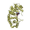
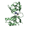
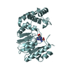
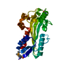
- PDB-7aye: Crystal structure of the computationally designed chemically disr... -
+
データを開く
IDまたはキーワード:
読み込み中...
-
基本情報
登録情報
データベース: PDB / ID: 7aye
タイトル
Crystal structure of the computationally designed chemically disruptable heterodimer LD6-MDM2
要素
Isoform 11 of E3 ubiquitin-protein ligase Mdm2
Thiol:disulfide interchange protein DsbD
キーワード
PROTEIN BINDING (タンパク質) / chemically disruptable heterodimer (CDH) / protein-protein interaction (タンパク質間相互作用) / gene and cell therapy / protein switches
機能・相同性
機能・相同性情報
タンパク質ジスルフィドレダクターゼ / protein-disulfide reductase (NAD(P)H) activity / cytochrome complex assembly / cellular response to vitamin B1 / response to formaldehyde / response to water-immersion restraint stress / traversing start control point of mitotic cell cycle / negative regulation of intrinsic apoptotic signaling pathway by p53 class mediator / response to ether / negative regulation of signal transduction by p53 class mediator ...タンパク質ジスルフィドレダクターゼ / protein-disulfide reductase (NAD(P)H) activity / cytochrome complex assembly / cellular response to vitamin B1 / response to formaldehyde / response to water-immersion restraint stress / traversing start control point of mitotic cell cycle / negative regulation of intrinsic apoptotic signaling pathway by p53 class mediator / response to ether / negative regulation of signal transduction by p53 class mediator / fibroblast activation / atrial septum development / receptor serine/threonine kinase binding / Trafficking of AMPA receptors / positive regulation of vascular associated smooth muscle cell migration / peroxisome proliferator activated receptor binding / response to iron ion / negative regulation of protein processing / SUMO transferase activity / response to steroid hormone / NEDD8 ligase activity / AKT phosphorylates targets in the cytosol / atrioventricular valve morphogenesis / cellular response to peptide hormone stimulus / ventricular septum development / endocardial cushion morphogenesis / positive regulation of muscle cell differentiation / SUMOylation of ubiquitinylation proteins / cellular response to alkaloid / blood vessel development / regulation of protein catabolic process / cardiac septum morphogenesis / Constitutive Signaling by AKT1 E17K in Cancer / ligase activity / negative regulation of DNA damage response, signal transduction by p53 class mediator / response to magnesium ion / protein sumoylation / SUMOylation of transcription factors / protein localization to nucleus / cellular response to UV-C / blood vessel remodeling / cellular response to estrogen stimulus / protein autoubiquitination / cellular response to actinomycin D / ribonucleoprotein complex binding / positive regulation of vascular associated smooth muscle cell proliferation / DNA damage response, signal transduction by p53 class mediator resulting in cell cycle arrest / NPAS4 regulates expression of target genes / transcription repressor complex / regulation of heart rate / positive regulation of mitotic cell cycle / proteolysis involved in protein catabolic process / positive regulation of protein export from nucleus / response to cocaine / ubiquitin binding / Stabilization of p53 / Regulation of RUNX3 expression and activity / protein destabilization / RING-type E3 ubiquitin transferase / Signaling by ALK fusions and activated point mutants / establishment of protein localization / Oncogene Induced Senescence / Regulation of TP53 Activity through Methylation / cellular response to gamma radiation / response to toxic substance / cellular response to growth factor stimulus / cellular response to hydrogen peroxide / protein polyubiquitination / ubiquitin-protein transferase activity / endocytic vesicle membrane / ubiquitin protein ligase activity / disordered domain specific binding / Regulation of TP53 Degradation / positive regulation of proteasomal ubiquitin-dependent protein catabolic process / p53 binding / negative regulation of neuron projection development / 5S rRNA binding / cellular response to hypoxia / ubiquitin-dependent protein catabolic process / 遺伝子発現の調節 / proteasome-mediated ubiquitin-dependent protein catabolic process / protein-containing complex assembly / Oxidative Stress Induced Senescence / Regulation of TP53 Activity through Phosphorylation / amyloid fibril formation / membrane => GO:0016020 / electron transfer activity / Ub-specific processing proteases / protein ubiquitination / regulation of cell cycle / response to xenobiotic stimulus / protein domain specific binding / response to antibiotic / negative regulation of DNA-templated transcription / apoptotic process / ubiquitin protein ligase binding / positive regulation of cell population proliferation / positive regulation of gene expression / 核小体 / negative regulation of apoptotic process 類似検索 - 分子機能
Thiol:disulfide interchange protein DsbD, N-terminal domain superfamily / Thiol:disulphide interchange protein DsbD / DsbD gamma / Thiol:disulfide interchange protein DsbD, N-terminal domain / Thiol:disulfide interchange protein DsbD, N-terminal / Cytochrome C biogenesis protein, transmembrane domain / Cytochrome C biogenesis protein transmembrane region / E3 ubiquitin-protein ligase Mdm2 / MDM2, modified RING finger, HC subclass / p53 negative regulator Mdm2/Mdm4 ...Thiol:disulfide interchange protein DsbD, N-terminal domain superfamily / Thiol:disulphide interchange protein DsbD / DsbD gamma / Thiol:disulfide interchange protein DsbD, N-terminal domain / Thiol:disulfide interchange protein DsbD, N-terminal / Cytochrome C biogenesis protein, transmembrane domain / Cytochrome C biogenesis protein transmembrane region / E3 ubiquitin-protein ligase Mdm2 / MDM2, modified RING finger, HC subclass / p53 negative regulator Mdm2/Mdm4 / SWIB/MDM2 domain / SWIB/MDM2 domain / SWIB/MDM2 domain profile. / SWIB/MDM2 domain superfamily / Zn-finger in Ran binding protein and others / Zinc finger, C3HC4 type (RING finger) / Zinc finger RanBP2 type profile. / Zinc finger RanBP2-type signature. / Zinc finger, RanBP2-type superfamily / Zinc finger, RanBP2-type / Thioredoxin, conserved site / Thioredoxin family active site. / Thioredoxin domain profile. / Thioredoxin domain / Zinc finger RING-type profile. / Zinc finger, RING-type / Thioredoxin-like superfamily / Zinc finger, RING/FYVE/PHD-type 類似検索 - ドメイン・相同性
解像度: 2.95→45.2 Å / Cor.coef. Fo:Fc: 0.949 / Cor.coef. Fo:Fc free: 0.931 / SU B: 21.452 / SU ML: 0.383 / 交差検証法: THROUGHOUT / σ(F): 0 / ESU R Free: 0.434 / 立体化学のターゲット値: MAXIMUM LIKELIHOOD 詳細: HYDROGENS HAVE BEEN ADDED IN THE RIDING POSITIONS U VALUES : REFINED INDIVIDUALLY
Rfactor
反射数
%反射
Selection details
Rfree
0.2599
285
5 %
RANDOM
Rwork
0.1971
-
-
-
obs
0.2006
5361
99.81 %
-
溶媒の処理
イオンプローブ半径: 0.8 Å / 減衰半径: 0.8 Å / VDWプローブ半径: 1.2 Å / 溶媒モデル: MASK
 ムービー
ムービー コントローラー


 データを開く
 基本情報
基本情報 要素
要素 キーワード
キーワード PROTEIN BINDING (タンパク質) / chemically disruptable heterodimer (CDH) /
  機能・相同性情報

 データ登録者
 引用
引用 構造の表示
構造の表示 ダウンロードとリンク
 その他のダウンロード
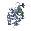


 PDBj
PDBj


 集合体
集合体
 試料調製
試料調製 / ビームライン: X06DA / 波長: 1 Å
 解析
解析