 Movie
Movie Controller

[English] 日本語
 Yorodumi
- PDB-7aye: Crystal structure of the computationally designed chemically disr... -
+ Open data

- Basic information
| Entry | Database: PDB / ID: 7aye | ||||||
|---|---|---|---|---|---|---|---|
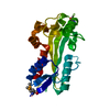
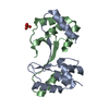
| Title | Crystal structure of the computationally designed chemically disruptable heterodimer LD6-MDM2 | ||||||
 Components Components |
| ||||||
 Keywords Keywords | PROTEIN BINDING / chemically disruptable heterodimer (CDH) / protein-protein interaction / gene and cell therapy / protein switches | ||||||
| Function / homology |  Function and homology information Function and homology informationprotein-disulfide reductase / protein-disulfide reductase [NAD(P)H] activity / cellular response to vitamin B1 / response to formaldehyde / cytochrome complex assembly / response to water-immersion restraint stress / response to ether / traversing start control point of mitotic cell cycle / atrial septum development / regulation of protein catabolic process at postsynapse, modulating synaptic transmission ...protein-disulfide reductase / protein-disulfide reductase [NAD(P)H] activity / cellular response to vitamin B1 / response to formaldehyde / cytochrome complex assembly / response to water-immersion restraint stress / response to ether / traversing start control point of mitotic cell cycle / atrial septum development / regulation of protein catabolic process at postsynapse, modulating synaptic transmission / fibroblast activation / Trafficking of AMPA receptors / receptor serine/threonine kinase binding / peroxisome proliferator activated receptor binding / negative regulation of intrinsic apoptotic signaling pathway by p53 class mediator / positive regulation of vascular associated smooth muscle cell migration / negative regulation of protein processing / SUMO transferase activity / response to steroid hormone / AKT phosphorylates targets in the cytosol / response to iron ion / NEDD8 ligase activity / atrioventricular valve morphogenesis / cellular response to peptide hormone stimulus / endocardial cushion morphogenesis / ventricular septum development / positive regulation of muscle cell differentiation / cardiac septum morphogenesis / regulation of postsynaptic neurotransmitter receptor internalization / blood vessel development / SUMOylation of ubiquitinylation proteins / ligase activity / cellular response to alkaloid / Constitutive Signaling by AKT1 E17K in Cancer / regulation of protein catabolic process / negative regulation of signal transduction by p53 class mediator / negative regulation of DNA damage response, signal transduction by p53 class mediator / SUMOylation of transcription factors / response to magnesium ion / cellular response to UV-C / protein sumoylation / cellular response to actinomycin D / cellular response to estrogen stimulus / blood vessel remodeling / protein localization to nucleus / ribonucleoprotein complex binding / protein autoubiquitination / positive regulation of vascular associated smooth muscle cell proliferation / NPAS4 regulates expression of target genes / transcription repressor complex / positive regulation of mitotic cell cycle / regulation of heart rate / proteolysis involved in protein catabolic process / positive regulation of protein export from nucleus / ubiquitin binding / response to cocaine / DNA damage response, signal transduction by p53 class mediator / Stabilization of p53 / establishment of protein localization / Regulation of RUNX3 expression and activity / cellular response to gamma radiation / Oncogene Induced Senescence / RING-type E3 ubiquitin transferase / protein destabilization / Regulation of TP53 Activity through Methylation / cellular response to growth factor stimulus / response to toxic substance / centriolar satellite / cellular response to hydrogen peroxide / protein polyubiquitination / ubiquitin-protein transferase activity / disordered domain specific binding / p53 binding / ubiquitin protein ligase activity / endocytic vesicle membrane / Signaling by ALK fusions and activated point mutants / Regulation of TP53 Degradation / positive regulation of proteasomal ubiquitin-dependent protein catabolic process / negative regulation of neuron projection development / 5S rRNA binding / protein-containing complex assembly / ubiquitin-dependent protein catabolic process / Oxidative Stress Induced Senescence / cellular response to hypoxia / Regulation of TP53 Activity through Phosphorylation / amyloid fibril formation / proteasome-mediated ubiquitin-dependent protein catabolic process / electron transfer activity / regulation of cell cycle / Ub-specific processing proteases / postsynaptic density / protein ubiquitination / response to xenobiotic stimulus / protein domain specific binding / response to antibiotic / negative regulation of DNA-templated transcription / positive regulation of cell population proliferation / apoptotic process / ubiquitin protein ligase binding / positive regulation of gene expression Similarity search - Function | ||||||
| Biological species |  Homo sapiens (human) Homo sapiens (human) Shigella dysenteriae Sd197 (bacteria) Shigella dysenteriae Sd197 (bacteria) | ||||||
| Method |  X-RAY DIFFRACTION / SYNCHROTRON / MOLECULAR REPLACEMENT / Resolution: 2.95 Å X-RAY DIFFRACTION / SYNCHROTRON / MOLECULAR REPLACEMENT / Resolution: 2.95 Å | ||||||
 Authors Authors | Yang, C. / Lau, K. / Pojer, F. / Correia, B.E. | ||||||
| Funding support | European Union, 1items
| ||||||
 Citation Citation | Journal: Nat Commun / Year: 2021 Title: A rational blueprint for the design of chemically-controlled protein switches. Authors: Shui, S. / Gainza, P. / Scheller, L. / Yang, C. / Kurumida, Y. / Rosset, S. / Georgeon, S. / Di Roberto, R.B. / Castellanos-Rueda, R. / Reddy, S.T. / Correia, B.E. | ||||||
| History |
|
- Structure visualization
| Structure viewer | Molecule: MolmilJmol/JSmol |
|---|
- Downloads & links
-Download
| PDBx/mmCIF format | 7aye.cif.gz | 56.3 KB | Display | PDBx/mmCIF format |
|---|---|---|---|---|
| PDB format | pdb7aye.ent.gz | 38.8 KB | Display | PDB format |
| PDBx/mmJSON format | 7aye.json.gz | Tree view | PDBx/mmJSON format | |
| Others |  Other downloads Other downloads |
-Validation report
| Summary document | 7aye_validation.pdf.gz | 437.1 KB | Display | wwPDB validaton report |
|---|---|---|---|---|
| Full document | 7aye_full_validation.pdf.gz | 439 KB | Display | |
| Data in XML | 7aye_validation.xml.gz | 9.3 KB | Display | |
| Data in CIF | 7aye_validation.cif.gz | 11.6 KB | Display | |
| Arichive directory | https://data.pdbj.org/pub/pdb/validation_reports/ay/7ayeftp://data.pdbj.org/pub/pdb/validation_reports/ay/7aye | HTTPS FTP |
-Related structure data
| Related structure data |  5afgS S: Starting model for refinement |
|---|---|
| Similar structure data |


-Links
 PDBj
PDBj


- Assembly
| Deposited unit | 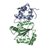
| ||||||||
|---|---|---|---|---|---|---|---|---|---|
| 1 |
| ||||||||
| Unit cell |
|
-Components
| #1: Protein | Mass: 13185.105 Da / Num. of mol.: 1 Source method: isolated from a genetically manipulated source Source: (gene. exp.) Homo sapiens (human) / Gene: MDM2 / Production host: References: UniProt: Q00987, RING-type E3 ubiquitin transferase |
|---|---|
| #2: Protein | Mass: 15537.526 Da / Num. of mol.: 1 Source method: isolated from a genetically manipulated source Source: (gene. exp.) Shigella dysenteriae Sd197 (bacteria) / Gene: dsbD, SDY_4441 / Production host: |
| Has protein modification | Y |
-Experimental details
-Experiment
| Experiment | Method: X-RAY DIFFRACTION / Number of used crystals: 1 |
|---|
- Sample preparation
| Crystal | Density Matthews: 2.15 Å3/Da / Density % sol: 42.89 % |
|---|---|
| Crystal grow | Temperature: 291 K / Method: vapor diffusion, sitting drop Details: 1.5 M Ammonium sulfate, 0.1 M Sodium cacodylate pH 6.5 |
-Data collection
| Diffraction | Mean temperature: 100 K / Serial crystal experiment: N |
|---|---|
| Diffraction source | Source: SYNCHROTRON / Site: SLS  / Beamline: X06DA / Wavelength: 1 Å / Beamline: X06DA / Wavelength: 1 Å |
| Detector | Type: DECTRIS PILATUS 2M-F / Detector: PIXEL / Date: Mar 2, 2018 |
| Radiation | Protocol: SINGLE WAVELENGTH / Monochromatic (M) / Laue (L): M / Scattering type: x-ray |
| Radiation wavelength | Wavelength: 1 Å / Relative weight: 1 |
| Reflection | Resolution: 2.95→45.2 Å / Num. obs: 5646 / % possible obs: 99.8 % / Redundancy: 6.9 % / CC1/2: 0.99 / Net I/σ(I): 22.18 |
| Reflection shell | Resolution: 2.95→3.13 Å / Mean I/σ(I) obs: 2.19 / Num. unique obs: 872 / CC1/2: 0.67 / % possible all: 99.8 |
- Processing
| Software |
| ||||||||||||||||||||||||||||||||||||||||||||||||||||||||||||
|---|---|---|---|---|---|---|---|---|---|---|---|---|---|---|---|---|---|---|---|---|---|---|---|---|---|---|---|---|---|---|---|---|---|---|---|---|---|---|---|---|---|---|---|---|---|---|---|---|---|---|---|---|---|---|---|---|---|---|---|---|---|
| Refinement | Method to determine structure: MOLECULAR REPLACEMENT Starting model: 5AFG Resolution: 2.95→45.2 Å / Cor.coef. Fo:Fc: 0.949 / Cor.coef. Fo:Fc free: 0.931 / SU B: 21.452 / SU ML: 0.383 / Cross valid method: THROUGHOUT / σ(F): 0 / ESU R Free: 0.434 / Stereochemistry target values: MAXIMUM LIKELIHOOD Details: HYDROGENS HAVE BEEN ADDED IN THE RIDING POSITIONS U VALUES : REFINED INDIVIDUALLY
| ||||||||||||||||||||||||||||||||||||||||||||||||||||||||||||
| Solvent computation | Ion probe radii: 0.8 Å / Shrinkage radii: 0.8 Å / VDW probe radii: 1.2 Å / Solvent model: MASK | ||||||||||||||||||||||||||||||||||||||||||||||||||||||||||||
| Displacement parameters | Biso max: 174.21 Å2 / Biso mean: 86.097 Å2 / Biso min: 52.11 Å2
| ||||||||||||||||||||||||||||||||||||||||||||||||||||||||||||
| Refinement step | Cycle: final / Resolution: 2.95→45.2 Å
| ||||||||||||||||||||||||||||||||||||||||||||||||||||||||||||
| Refine LS restraints |
| ||||||||||||||||||||||||||||||||||||||||||||||||||||||||||||
| LS refinement shell | Resolution: 2.954→3.031 Å / Rfactor Rfree error: 0 / Total num. of bins used: 20
|
