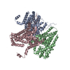
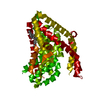
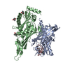
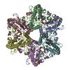
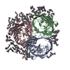
Journal: EMBO J / Year: 2022 Title: The ion-coupling mechanism of human excitatory amino acid transporters. Authors: Juan C Canul-Tec / Anand Kumar / Jonathan Dhenin / Reda Assal / Pierre Legrand / Martial Rey / Julia Chamot-Rooke / Nicolas Reyes / Abstract: Excitatory amino acid transporters (EAATs) maintain glutamate gradients in the brain essential for neurotransmission and to prevent neuronal death. They use ionic gradients as energy source and co- ...Excitatory amino acid transporters (EAATs) maintain glutamate gradients in the brain essential for neurotransmission and to prevent neuronal death. They use ionic gradients as energy source and co-transport transmitter into the cytoplasm with Na and H , while counter-transporting K to re-initiate the transport cycle. However, the molecular mechanisms underlying ion-coupled transport remain incompletely understood. Here, we present 3D X-ray crystallographic and cryo-EM structures, as well as thermodynamic analysis of human EAAT1 in different ion bound conformations, including elusive counter-transport ion bound states. Binding energies of Na and H , and unexpectedly Ca , are coupled to neurotransmitter binding. Ca competes for a conserved Na site, suggesting a regulatory role for Ca in glutamate transport at the synapse, while H binds to a conserved glutamate residue stabilizing substrate occlusion. The counter-transported ion binding site overlaps with that of glutamate, revealing the K -based mechanism to exclude the transmitter during the transport cycle and to prevent its neurotoxic release on the extracellular side.
In the structure databanks used in Yorodumi, some data are registered as the other names, "COVID-19 virus" and "2019-nCoV". Here are the details of the virus and the list of structure data.
Jan 31, 2019. EMDB accession codes are about to change! (news from PDBe EMDB page)
EMDB accession codes are about to change! (news from PDBe EMDB page)
The allocation of 4 digits for EMDB accession codes will soon come to an end. Whilst these codes will remain in use, new EMDB accession codes will include an additional digit and will expand incrementally as the available range of codes is exhausted. The current 4-digit format prefixed with “EMD-” (i.e. EMD-XXXX) will advance to a 5-digit format (i.e. EMD-XXXXX), and so on. It is currently estimated that the 4-digit codes will be depleted around Spring 2019, at which point the 5-digit format will come into force.
The EM Navigator/Yorodumi systems omit the EMD- prefix.
Related info.:Q: What is EMD? / ID/Accession-code notation in Yorodumi/EM Navigator
Yorodumi is a browser for structure data from EMDB, PDB, SASBDB, etc.
This page is also the successor to EM Navigator detail page, and also detail information page/front-end page for Omokage search.
The word "yorodu" (or yorozu) is an old Japanese word meaning "ten thousand". "mi" (miru) is to see.
Related info.:EMDB / PDB / SASBDB / Comparison of 3 databanks / Yorodumi Search / Aug 31, 2016. New EM Navigator & Yorodumi / Yorodumi Papers / Jmol/JSmol / Function and homology information / Changes in new EM Navigator and Yorodumi
 Movie
Movie Controller

 Yorodumi
 Open data
 Basic information
 Components
 Keywords
 Function and homology information
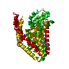
 Homo sapiens (human)
 X-RAY DIFFRACTION /
  Authors
 France, 1items
  Citation
 Structure visualization
 Downloads & links
 Other downloads


 PDBj
PDBj Assembly
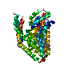
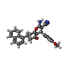
 Mass: 422.475 Da / Num. of mol.: 1 / Source method: obtained synthetically / Formula: C27H22N2O3
 Mass: 137.327 Da / Num. of mol.: 2 / Source method: obtained synthetically / Formula: Ba / Feature type: SUBJECT OF INVESTIGATION
 Sample preparation
 Processing