 Movie
Movie Controller

[English] 日本語
 Yorodumi
- PDB-2po1: Crystal structure of the P. abyssi exosome RNase PH ring complexe... -
+ Open data

- Basic information
| Entry | Database: PDB / ID: 2po1 | ||||||
|---|---|---|---|---|---|---|---|
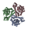
| Title | Crystal structure of the P. abyssi exosome RNase PH ring complexed with a single stranded 10-mer poly(A) RNA | ||||||
 Components Components |
| ||||||
 Keywords Keywords | hydrolase/hydrolase/RNA / RNase PH / hydrolase-hydrolase-RNA COMPLEX | ||||||
| Function / homology |  Function and homology information Function and homology informationcytoplasmic exosome (RNase complex) / rRNA catabolic process / mRNA 3'-UTR AU-rich region binding / nuclear-transcribed mRNA catabolic process / Hydrolases; Acting on ester bonds; Exoribonucleases producing 5'-phosphomonoesters / 3'-5'-RNA exonuclease activity / gene expression / RNA binding Similarity search - Function | ||||||
| Biological species |   Pyrococcus abyssi (archaea) Pyrococcus abyssi (archaea) | ||||||
| Method |  X-RAY DIFFRACTION / SYNCHROTRON / MOLECULAR REPLACEMENT / Resolution: 1.94 Å X-RAY DIFFRACTION / SYNCHROTRON / MOLECULAR REPLACEMENT / Resolution: 1.94 Å | ||||||
 Authors Authors | Navarro, M.V.A.S. / Guimaraes, B.G. | ||||||
 Citation Citation | Journal: J.Biol.Chem. / Year: 2008 Title: Insights into the mechanism of progressive RNA degradation by the archaeal exosome. Authors: Navarro, M.V.A.S. / Oliveira, C.C. / Zanchin, N.I. / Guimaraes, B.G. | ||||||
| History |
|
- Structure visualization
| Structure viewer | Molecule: MolmilJmol/JSmol |
|---|
- Downloads & links
-Download
| PDBx/mmCIF format | 2po1.cif.gz | 231.8 KB | Display | PDBx/mmCIF format |
|---|---|---|---|---|
| PDB format | pdb2po1.ent.gz | 185.9 KB | Display | PDB format |
| PDBx/mmJSON format | 2po1.json.gz | Tree view | PDBx/mmJSON format | |
| Others |  Other downloads Other downloads |
-Validation report
| Arichive directory | https://data.pdbj.org/pub/pdb/validation_reports/po/2po1ftp://data.pdbj.org/pub/pdb/validation_reports/po/2po1 | HTTPS FTP |
|---|
-Related structure data
| Related structure data | 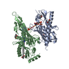 2pnzSC 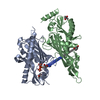 2po0C 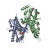 2po2C S: Starting model for refinement C: citing same article ( |
|---|---|
| Similar structure data |


-Links
 PDBj
PDBj
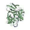
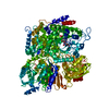
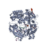
- Assembly
| Deposited unit | 
| ||||||||
|---|---|---|---|---|---|---|---|---|---|
| 1 | 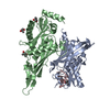
| ||||||||
| Unit cell |
| ||||||||
| Details | The biological assembly is a hexamer generated from the heterodimer in the asymmetric unit by the operations: -y, x-y-1, z and -x+y+1, -x, z |
-Components
| #1: RNA chain | Mass: 3247.100 Da / Num. of mol.: 1 / Source method: obtained synthetically | ||
|---|---|---|---|
| #2: Protein | Mass: 27720.305 Da / Num. of mol.: 1 Source method: isolated from a genetically manipulated source Source: (gene. exp.) Pyrococcus abyssi (archaea) / Gene: Rrp41 / Plasmid: pET29 / Species (production host): Escherichia coli / Production host:  References: UniProt: Q9V119, Hydrolases; Acting on ester bonds; Exoribonucleases producing 5'-phosphomonoesters | ||
| #3: Protein | Mass: 30268.832 Da / Num. of mol.: 1 Source method: isolated from a genetically manipulated source Source: (gene. exp.) Pyrococcus abyssi (archaea) / Gene: Rrp42 / Plasmid: pAE / Species (production host): Escherichia coli / Production host: References: UniProt: Q9V118, Hydrolases; Acting on ester bonds; Exoribonucleases producing 5'-phosphomonoesters | ||
| #4: Chemical | ChemComp-MPD / (   Mass: 118.174 Da / Num. of mol.: 5 / Source method: obtained synthetically / Formula: C6H14O2 / Comment: precipitant*YM Mass: 118.174 Da / Num. of mol.: 5 / Source method: obtained synthetically / Formula: C6H14O2 / Comment: precipitant*YM#5: Water | ChemComp-HOH / |  Mass: 18.015 Da / Num. of mol.: 308 / Source method: isolated from a natural source / Formula: H2O Mass: 18.015 Da / Num. of mol.: 308 / Source method: isolated from a natural source / Formula: H2O |
-Experimental details
-Experiment
| Experiment | Method: X-RAY DIFFRACTION / Number of used crystals: 1 |
|---|
- Sample preparation
| Crystal | Density Matthews: 2.61 Å3/Da / Density % sol: 52.86 % | ||||||||||||||||||||||||||||
|---|---|---|---|---|---|---|---|---|---|---|---|---|---|---|---|---|---|---|---|---|---|---|---|---|---|---|---|---|---|
| Crystal grow | Temperature: 291 K / Method: vapor diffusion, hanging drop / pH: 6 Details: 0.1 M Bis-Tris, 45% MPD and 0.1 M LiCl, pH 6.0, VAPOR DIFFUSION, HANGING DROP, temperature 291.0K | ||||||||||||||||||||||||||||
| Components of the solutions |
|
-Data collection
| Diffraction | Mean temperature: 100 K |
|---|---|
| Diffraction source | Source: SYNCHROTRON / Site: LNLS  / Beamline: D03B-MX1 / Wavelength: 1.427 Å / Beamline: D03B-MX1 / Wavelength: 1.427 Å |
| Detector | Type: MAR CCD 165 mm / Detector: CCD / Date: May 10, 2006 / Details: mirrors |
| Radiation | Monochromator: Si 111 CHANNEL / Protocol: SINGLE WAVELENGTH / Monochromatic (M) / Laue (L): M / Scattering type: x-ray |
| Radiation wavelength | Wavelength: 1.427 Å / Relative weight: 1 |
| Reflection | Resolution: 1.94→46.78 Å / Num. all: 47831 / Num. obs: 46981 / % possible obs: 98.2 % / Observed criterion σ(F): 1 / Observed criterion σ(I): 1 / Redundancy: 11 % / Biso Wilson estimate: 23.6 Å2 / Rsym value: 0.109 / Net I/σ(I): 14.9 |
| Reflection shell | Resolution: 1.94→2.06 Å / Redundancy: 9.2 % / Mean I/σ(I) obs: 3.5 / Num. unique all: 6859 / Rsym value: 0.627 / % possible all: 90.1 |
- Processing
| Software |
| |||||||||||||||||||||||||||||||||||||||||||||||||||||||||||||||||||||||||||||||||||||||||||||||||||||||||||||||||||||||||||||
|---|---|---|---|---|---|---|---|---|---|---|---|---|---|---|---|---|---|---|---|---|---|---|---|---|---|---|---|---|---|---|---|---|---|---|---|---|---|---|---|---|---|---|---|---|---|---|---|---|---|---|---|---|---|---|---|---|---|---|---|---|---|---|---|---|---|---|---|---|---|---|---|---|---|---|---|---|---|---|---|---|---|---|---|---|---|---|---|---|---|---|---|---|---|---|---|---|---|---|---|---|---|---|---|---|---|---|---|---|---|---|---|---|---|---|---|---|---|---|---|---|---|---|---|---|---|---|
| Refinement | Method to determine structure: MOLECULAR REPLACEMENT Starting model: PDB ENTRY 2PNZ Resolution: 1.94→20 Å / Cor.coef. Fo:Fc: 0.956 / Cor.coef. Fo:Fc free: 0.927 / SU B: 9.473 / SU ML: 0.122 / Isotropic thermal model: ISOTROPIC / Cross valid method: THROUGHOUT / σ(F): 1 / σ(I): 1 / ESU R: 0.151 / ESU R Free: 0.152 / Stereochemistry target values: MAXIMUM LIKELIHOOD / Details: HYDROGENS HAVE BEEN ADDED IN THE RIDING POSITIONS
| |||||||||||||||||||||||||||||||||||||||||||||||||||||||||||||||||||||||||||||||||||||||||||||||||||||||||||||||||||||||||||||
| Solvent computation | Ion probe radii: 0.8 Å / Shrinkage radii: 0.8 Å / VDW probe radii: 1.4 Å / Solvent model: MASK | |||||||||||||||||||||||||||||||||||||||||||||||||||||||||||||||||||||||||||||||||||||||||||||||||||||||||||||||||||||||||||||
| Displacement parameters | Biso mean: 29.413 Å2
| |||||||||||||||||||||||||||||||||||||||||||||||||||||||||||||||||||||||||||||||||||||||||||||||||||||||||||||||||||||||||||||
| Refinement step | Cycle: LAST / Resolution: 1.94→20 Å
| |||||||||||||||||||||||||||||||||||||||||||||||||||||||||||||||||||||||||||||||||||||||||||||||||||||||||||||||||||||||||||||
| Refine LS restraints |
| |||||||||||||||||||||||||||||||||||||||||||||||||||||||||||||||||||||||||||||||||||||||||||||||||||||||||||||||||||||||||||||
| LS refinement shell | Resolution: 1.94→1.99 Å / Total num. of bins used: 20
| |||||||||||||||||||||||||||||||||||||||||||||||||||||||||||||||||||||||||||||||||||||||||||||||||||||||||||||||||||||||||||||
| Refinement TLS params. | Method: refined / Refine-ID: X-RAY DIFFRACTION
| |||||||||||||||||||||||||||||||||||||||||||||||||||||||||||||||||||||||||||||||||||||||||||||||||||||||||||||||||||||||||||||
| Refinement TLS group |
|
