 Movie
Movie Controller


+ Open data

- Basic information
| Entry | Database: PDB / ID: 7avm | ||||||
|---|---|---|---|---|---|---|---|
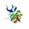
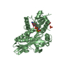
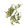
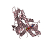
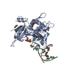
| Title | Crystal Structure of Pro-Rhodesain C150A | ||||||
 Components Components | Cysteine protease | ||||||
 Keywords Keywords | HYDROLASE / African trypanosomes / Sleeping Sickness / human african trypanosomiasis / cysteine protease / zymogen / pro-enzyme / rhodesain | ||||||
| Function / homology |  Function and homology information Function and homology information | ||||||
| Biological species |  | ||||||
| Method |  X-RAY DIFFRACTION / MOLECULAR REPLACEMENT / Resolution: 2.8 Å X-RAY DIFFRACTION / MOLECULAR REPLACEMENT / Resolution: 2.8 Å | ||||||
 Authors Authors | Johe, P. / Jaenicke, E. / Neuweiler, H. / Schirmeister, T. / Kersten, C. / Hellmich, U. | ||||||
 Citation Citation | Journal: J.Biol.Chem. / Year: 2021 Title: Structure, interdomain dynamics, and pH-dependent autoactivation of pro-rhodesain, the main lysosomal cysteine protease from African trypanosomes. Authors: Johe, P. / Jaenicke, E. / Neuweiler, H. / Schirmeister, T. / Kersten, C. / Hellmich, U.A. #1: Journal: Biorxiv / Year: 2020Title: Structural basis of autoinhibition in the T. brucei rhodesiense cathepsin L zymogen pro-rhodesain and pH-dependent cleavage Authors: Johe, P. / Jaenicke, E. / Neuweiler, H. / Schirmeister, T. / Kersten, C. / Hellmich, U. #2: Journal: To Be PublishedTitle: Structural basis of autoinhibition in the T. brucei rhodesiense cathepsin L zymogen pro-rhodesain and pH-dependent cleavage Authors: Johe, P. / Kersten, C. / Jaenicke, E. / Neuweiler, H. / Schirmeister, T. / Hellmich, U. | ||||||
| History |
|
- Structure visualization
| Structure viewer | Molecule: MolmilJmol/JSmol |
|---|
- Downloads & links
-Download
| PDBx/mmCIF format | 7avm.cif.gz | 76.4 KB | Display | PDBx/mmCIF format |
|---|---|---|---|---|
| PDB format | pdb7avm.ent.gz | 54 KB | Display | PDB format |
| PDBx/mmJSON format | 7avm.json.gz | Tree view | PDBx/mmJSON format | |
| Others |  Other downloads Other downloads |
-Validation report
| Arichive directory | https://data.pdbj.org/pub/pdb/validation_reports/av/7avmftp://data.pdbj.org/pub/pdb/validation_reports/av/7avm | HTTPS FTP |
|---|
-Related structure data
| Related structure data |  6ex8S S: Starting model for refinement |
|---|---|
| Similar structure data |


-Links
 PDBj
PDBj


- Assembly
| Deposited unit | 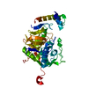
| ||||||||||||
|---|---|---|---|---|---|---|---|---|---|---|---|---|---|
| 1 |
| ||||||||||||
| Unit cell |
|
-Components
| #1: Protein | Mass: 35897.926 Da / Num. of mol.: 1 / Mutation: C150A Source method: isolated from a genetically manipulated source Source: (gene. exp.) Gene: rhodesain / Production host:  |
|---|---|
| #2: Chemical | ChemComp-GOL /   Mass: 92.094 Da / Num. of mol.: 1 / Source method: obtained synthetically / Formula: C3H8O3 Mass: 92.094 Da / Num. of mol.: 1 / Source method: obtained synthetically / Formula: C3H8O3 |
| #3: Water | ChemComp-HOH /  Mass: 18.015 Da / Num. of mol.: 38 / Source method: isolated from a natural source / Formula: H2O Mass: 18.015 Da / Num. of mol.: 38 / Source method: isolated from a natural source / Formula: H2O |
| Has ligand of interest | N |
| Has protein modification | Y |
-Experimental details
-Experiment
| Experiment | Method: X-RAY DIFFRACTION / Number of used crystals: 1 |
|---|
- Sample preparation
| Crystal | Density Matthews: 2.07 Å3/Da / Density % sol: 40.48 % |
|---|---|
| Crystal grow | Temperature: 300 K / Method: vapor diffusion, hanging drop / pH: 3.5 Details: 40 mM sodium citrate, 30% PEG-6000, Lead(II) acetate (saturated),cryoprotection with glycerol 10% |
-Data collection
| Diffraction | Mean temperature: 100 K / Serial crystal experiment: N |
|---|---|
| Diffraction source | Source: ROTATING ANODE / Type: BRUKER AXS MICROSTAR-H / Wavelength: 1.5417 Å |
| Detector | Type: MAR scanner 300 mm plate / Detector: IMAGE PLATE / Date: Jul 11, 2018 |
| Radiation | Protocol: SINGLE WAVELENGTH / Monochromatic (M) / Laue (L): M / Scattering type: x-ray |
| Radiation wavelength | Wavelength: 1.5417 Å / Relative weight: 1 |
| Reflection | Resolution: 2.8→37.92 Å / Num. obs: 7512 / % possible obs: 97.23 % / Redundancy: 20.6 % / Biso Wilson estimate: 41.24 Å2 / CC1/2: 0.919 / CC star: 0.979 / Net I/σ(I): 214.7 |
| Reflection shell | Resolution: 2.8→2.9 Å / Num. unique obs: 729 / CC1/2: 0.905 / CC star: 0.975 / % possible all: 92.73 |
- Processing
| Software |
| ||||||||||||||||||||||||||||||||||||||||||
|---|---|---|---|---|---|---|---|---|---|---|---|---|---|---|---|---|---|---|---|---|---|---|---|---|---|---|---|---|---|---|---|---|---|---|---|---|---|---|---|---|---|---|---|
| Refinement | Method to determine structure: MOLECULAR REPLACEMENT Starting model: 6EX8 Resolution: 2.8→37.92 Å / SU ML: 0.265 / Cross valid method: FREE R-VALUE / σ(F): 1.97 / Phase error: 28.3682 Stereochemistry target values: GeoStd + Monomer Library + CDL v1.2
| ||||||||||||||||||||||||||||||||||||||||||
| Solvent computation | Shrinkage radii: 0.9 Å / VDW probe radii: 1.11 Å / Solvent model: FLAT BULK SOLVENT MODEL | ||||||||||||||||||||||||||||||||||||||||||
| Displacement parameters | Biso mean: 47.97 Å2 | ||||||||||||||||||||||||||||||||||||||||||
| Refinement step | Cycle: LAST / Resolution: 2.8→37.92 Å
| ||||||||||||||||||||||||||||||||||||||||||
| Refine LS restraints |
| ||||||||||||||||||||||||||||||||||||||||||
| LS refinement shell |
|
