 ムービー
ムービー コントローラー


+ データを開く

- 基本情報
基本情報
| 登録情報 | データベース: PDB / ID: 7asi | ||||||||||||
|---|---|---|---|---|---|---|---|---|---|---|---|---|---|
| タイトル | Fixed-target serial femtosecond crystallography using in cellulo grown Neurospora crassa HEX-1 microcrystals. (Chips 1+2) | ||||||||||||
 要素 要素 | eIF-5a domain-containing protein | ||||||||||||
 キーワード キーワード | STRUCTURAL PROTEIN / in cellulo crystals / Woronin body / septal pore sealing | ||||||||||||
| 機能・相同性 |  機能・相同性情報 機能・相同性情報positive regulation of translational termination / positive regulation of translational elongation / translation elongation factor activity / ribosome binding / RNA binding 類似検索 - 分子機能 | ||||||||||||
| 生物種 |  Neurospora crassa (菌類) Neurospora crassa (菌類) | ||||||||||||
| 手法 |  X線回折 / 自由電子レーザー / 分子置換 / 解像度: 1.704 Å X線回折 / 自由電子レーザー / 分子置換 / 解像度: 1.704 Å | ||||||||||||
| Model details | Chip 1+2 | ||||||||||||
 データ登録者 データ登録者 | Lahey-Rudolph, J.M. / Schoenherr, R. / Barthelmess, M. / Fischer, P. / Seuring, C. / Wagner, A. / Meents, A. / Redecke, L. | ||||||||||||
| 資金援助 |  ドイツ, 3件 ドイツ, 3件
| ||||||||||||
 引用 引用 | ジャーナル: Iucrj / 年: 2021 タイトル: Fixed-target serial femtosecond crystallography using in cellulo grown microcrystals. 著者: Lahey-Rudolph, J.M. / Schonherr, R. / Barthelmess, M. / Fischer, P. / Seuring, C. / Wagner, A. / Meents, A. / Redecke, L. | ||||||||||||
| 履歴 |
|
- 構造の表示
構造の表示
| 構造ビューア | 分子: MolmilJmol/JSmol |
|---|
- ダウンロードとリンク
-ダウンロード
| PDBx/mmCIF形式 | 7asi.cif.gz | 68.6 KB | 表示 | PDBx/mmCIF形式 |
|---|---|---|---|---|
| PDB形式 | pdb7asi.ent.gz | 50.3 KB | 表示 | PDB形式 |
| PDBx/mmJSON形式 | 7asi.json.gz | ツリー表示 | PDBx/mmJSON形式 | |
| その他 |  その他のダウンロード その他のダウンロード |
-検証レポート
| 文書・要旨 | 7asi_validation.pdf.gz | 420.5 KB | 表示 | wwPDB検証レポート |
|---|---|---|---|---|
| 文書・詳細版 | 7asi_full_validation.pdf.gz | 431.4 KB | 表示 | |
| XML形式データ | 7asi_validation.xml.gz | 8.6 KB | 表示 | |
| CIF形式データ | 7asi_validation.cif.gz | 11.2 KB | 表示 | |
| アーカイブディレクトリ | https://data.pdbj.org/pub/pdb/validation_reports/as/7asiftp://data.pdbj.org/pub/pdb/validation_reports/as/7asi | HTTPS FTP |
-関連構造データ
| 関連構造データ |  7asxC 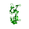 1khiS S: 精密化の開始モデル C: 同じ文献を引用 ( |
|---|---|
| 類似構造データ | |
| その他のデータベース |
|


-リンク
 PDBj
PDBj- 集合体
集合体
| 登録構造単位 | 
| ||||||||
|---|---|---|---|---|---|---|---|---|---|
| 1 | 
| ||||||||
| 単位格子 |
| ||||||||
| Components on special symmetry positions |
|
-要素
| #1: タンパク質 | 分子量: 19221.742 Da / 分子数: 1 / 変異: Met1_Tyr2insGly; Ser175_*insAla / 由来タイプ: 組換発現 詳細: Recombinant baculovirus production in Escherichia coli DH10EmBacY (Geneva Biotech, BacToBac system). Lipofection with Escort IV reagent. P3 recombinant baculovirus stock used for infection of ...詳細: Recombinant baculovirus production in Escherichia coli DH10EmBacY (Geneva Biotech, BacToBac system). Lipofection with Escort IV reagent. P3 recombinant baculovirus stock used for infection of insect cells at MOI 1. 由来: (組換発現) Neurospora crassa (菌類) / 遺伝子: GE21DRAFT_1527プラスミド: modified pFastBac1 vector containing the sequence 5 -ATGGGCGCCTAA-3 between the BamHI and HindIII restriction sites 細胞株 (発現宿主): Sf9 発現宿主:   Spodoptera frugiperda (ツマジロクサヨトウ) Spodoptera frugiperda (ツマジロクサヨトウ)参照: UniProt: A0A0B0EDT5 |
|---|---|
| #2: 水 | ChemComp-HOH /  分子量: 18.015 Da / 分子数: 94 / 由来タイプ: 天然 / 式: H2O 分子量: 18.015 Da / 分子数: 94 / 由来タイプ: 天然 / 式: H2O |
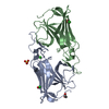
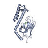
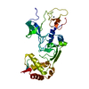
| 構成要素の詳細 | The in cellulo crystallized Neurospora crassa HEX-1 forms a polymeric helical spiral with 12 ...The in cellulo crystallized Neurospora crassa HEX-1 forms a polymeric helical spiral with 12 monomers per full turn, or 6 homodimeric units per full turn. The homodimeric assembly features an identical interaction on opposite faces that propagates the spiral. This results in the six-fold symmetry of the crystals that is consistently observed in native Woronin bodies |
-実験情報
-実験
| 実験 | 手法: X線回折 / 使用した結晶の数: 1 |
|---|
- 試料調製
試料調製
| 結晶 | マシュー密度: 2.51 Å3/Da / 溶媒含有率: 50.93 % |
|---|---|
| 結晶化 | 温度: 300.15 K / 手法: in cell 詳細: Neurospora crassa HEX-1 P3 rBV-infected Sf9 cells at multiplicity of infection 1, cultivated in serum-free ESF921 medium |
-データ収集
| 回折 | 平均測定温度: 298 K / Serial crystal experiment: Y |
|---|---|
| 放射光源 | 由来: 自由電子レーザー / サイト: SLAC LCLS  / ビームライン: MFX / 波長: 1.312 Å / ビームライン: MFX / 波長: 1.312 Å |
| 検出器 | タイプ: CS-PAD CXI-1 / 検出器: PIXEL / 日付: 2017年10月2日 / 詳細: Beryllium lenses |
| 放射 | プロトコル: SINGLE WAVELENGTH / 単色(M)・ラウエ(L): M / 散乱光タイプ: x-ray |
| 放射波長 | 波長: 1.312 Å / 相対比: 1 |
| 反射 | 解像度: 1.704→39.9 Å / Num. obs: 22635 / % possible obs: 99.99 % / 冗長度: 124.12 % / Biso Wilson estimate: 18.31 Å2 / CC1/2: 0.964 / CC star: 0.991 / R split: 0.162 / Net I/σ(I): 5.421 |
| 反射 シェル | 解像度: 1.704→1.744 Å / 冗長度: 16.4 % / Mean I/σ(I) obs: 0.7 / Num. unique obs: 1469 / CC1/2: 0.16 / CC star: 0.525 / R split: 0.189 / % possible all: 99.86 |
| Serial crystallography measurement | Collection time total: 0.25 hours / Focal spot size: 1 µm2 / Pulse duration: 27.5 fsec. / Pulse photon energy: 9.45 keV / Source distance: 0.116 m / XFEL pulse repetition rate: 120 Hz |
| Serial crystallography sample delivery | 解説: fixed target / 手法: fixed target |
| Serial crystallography sample delivery fixed target | 解説: micro-patterned single-crystalline silicon microchips 詳細: scanning speed matched to 120 Hz pulse repetition rate Motion control: attachment to Roadrunner II / Sample dehydration prevention: humidor, helium chamber / Sample solvent: ESF921 insect cell medium / Sample unit size: 12 µm / Support base: Chips glued to aluminium support frame / Velocity horizontal: 2.5 / Velocity vertical: 100 |
| Serial crystallography data reduction | Crystal hits: 19676 / Frames failed index: 6286 / Frames indexed: 13390 / Frames total: 87450 / Lattices indexed: 15224 / XFEL pulse events: 87450 / XFEL run numbers: 6,9 |
-位相決定
| 位相決定 | 手法: 分子置換 |
|---|
- 解析
解析
| ソフトウェア |
| ||||||||||||||||||||||||||||||||||||||||||||||||||||||||||||||||||||||||||||||
|---|---|---|---|---|---|---|---|---|---|---|---|---|---|---|---|---|---|---|---|---|---|---|---|---|---|---|---|---|---|---|---|---|---|---|---|---|---|---|---|---|---|---|---|---|---|---|---|---|---|---|---|---|---|---|---|---|---|---|---|---|---|---|---|---|---|---|---|---|---|---|---|---|---|---|---|---|---|---|---|
| 精密化 | 構造決定の手法: 分子置換 開始モデル: 1khi 解像度: 1.704→29.365 Å / SU ML: 0.29 / 交差検証法: THROUGHOUT / σ(F): 1.33 / 位相誤差: 22.95 / 立体化学のターゲット値: ML
| ||||||||||||||||||||||||||||||||||||||||||||||||||||||||||||||||||||||||||||||
| 溶媒の処理 | 減衰半径: 0.9 Å / VDWプローブ半径: 1.11 Å / 溶媒モデル: FLAT BULK SOLVENT MODEL | ||||||||||||||||||||||||||||||||||||||||||||||||||||||||||||||||||||||||||||||
| 原子変位パラメータ | Biso max: 87.52 Å2 / Biso mean: 25.3897 Å2 / Biso min: 3.79 Å2 | ||||||||||||||||||||||||||||||||||||||||||||||||||||||||||||||||||||||||||||||
| 精密化ステップ | サイクル: final / 解像度: 1.704→29.365 Å
| ||||||||||||||||||||||||||||||||||||||||||||||||||||||||||||||||||||||||||||||
| 拘束条件 |
| ||||||||||||||||||||||||||||||||||||||||||||||||||||||||||||||||||||||||||||||
| LS精密化 シェル | Refine-ID: X-RAY DIFFRACTION / Rfactor Rfree error: 0
|
