 Movie
Movie Controller

[English] 日本語
 Yorodumi
- PDB-7aqx: Co-Crystal Structure of Variant Surface Glycoprotein VSG2 in comp... -
+ Open data

- Basic information
| Entry | Database: PDB / ID: 7aqx | ||||||
|---|---|---|---|---|---|---|---|
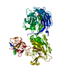
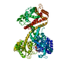
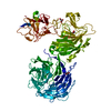
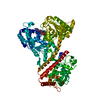
| Title | Co-Crystal Structure of Variant Surface Glycoprotein VSG2 in complex with Nanobody VSG2(NB9) | ||||||
 Components Components |
| ||||||
 Keywords Keywords | MEMBRANE PROTEIN / Variant surface Glycoprotein / Epitope Mapping / Immune Evasion | ||||||
| Function / homology |  Function and homology information Function and homology informationsymbiont-mediated evasion of host immune response / side of membrane / plasma membrane Similarity search - Function | ||||||
| Biological species |   | ||||||
| Method |  X-RAY DIFFRACTION / SYNCHROTRON / MOLECULAR REPLACEMENT / Resolution: 1.499 Å X-RAY DIFFRACTION / SYNCHROTRON / MOLECULAR REPLACEMENT / Resolution: 1.499 Å | ||||||
 Authors Authors | Stebbins, C.E. / Hempelmann, A. | ||||||
 Citation Citation | Journal: Cell Rep / Year: 2021 Title: Nanobody-mediated macromolecular crowding induces membrane fission and remodeling in the African trypanosome. Authors: Hempelmann, A. / Hartleb, L. / van Straaten, M. / Hashemi, H. / Zeelen, J.P. / Bongers, K. / Papavasiliou, F.N. / Engstler, M. / Stebbins, C.E. / Jones, N.G. | ||||||
| History |
|
- Structure visualization
| Structure viewer | Molecule: MolmilJmol/JSmol |
|---|
- Downloads & links
-Download
| PDBx/mmCIF format | 7aqx.cif.gz | 510.7 KB | Display | PDBx/mmCIF format |
|---|---|---|---|---|
| PDB format | pdb7aqx.ent.gz | 420.1 KB | Display | PDB format |
| PDBx/mmJSON format | 7aqx.json.gz | Tree view | PDBx/mmJSON format | |
| Others |  Other downloads Other downloads |
-Validation report
| Arichive directory | https://data.pdbj.org/pub/pdb/validation_reports/aq/7aqxftp://data.pdbj.org/pub/pdb/validation_reports/aq/7aqx | HTTPS FTP |
|---|
-Related structure data
| Related structure data |  7aqyC  7aqzC  7ar0C  1vsgS  5lhrS S: Starting model for refinement C: citing same article ( |
|---|---|
| Similar structure data |


-Links
 PDBj
PDBj

- Assembly
| Deposited unit | 
| ||||||||
|---|---|---|---|---|---|---|---|---|---|
| 1 |
| ||||||||
| Unit cell |
|
-Components
-Protein / Antibody , 2 types, 4 molecules ABCD
| #1: Protein | Mass: 38844.660 Da / Num. of mol.: 2 / Source method: isolated from a natural source / Details: Glycosylated at N289 / Source: (natural) #2: Antibody | Mass: 15722.019 Da / Num. of mol.: 2 Source method: isolated from a genetically manipulated source Details: Bound to Variant Surface Glycoprotein VSG2 / Source: (gene. exp.)  |
|---|
-Sugars , 2 types, 2 molecules
| #3: Polysaccharide | beta-D-mannopyranose-(1-4)-2-acetamido-2-deoxy-beta-D-glucopyranose-(1-4)-2-acetamido-2-deoxy-beta- ...beta-D-mannopyranose-(1-4)-2-acetamido-2-deoxy-beta-D-glucopyranose-(1-4)-2-acetamido-2-deoxy-beta-D-glucopyranose Source method: isolated from a genetically manipulated source |
|---|---|
| #4: Polysaccharide | alpha-D-mannopyranose-(1-3)-beta-D-mannopyranose-(1-4)-2-acetamido-2-deoxy-beta-D-glucopyranose-(1- ...alpha-D-mannopyranose-(1-3)-beta-D-mannopyranose-(1-4)-2-acetamido-2-deoxy-beta-D-glucopyranose-(1-4)-2-acetamido-2-deoxy-beta-D-glucopyranose Source method: isolated from a genetically manipulated source |
-Non-polymers , 3 types, 928 molecules 


| #5: Chemical | ChemComp-CIT /  Mass: 192.124 Da / Num. of mol.: 1 Mass: 192.124 Da / Num. of mol.: 1Source method: isolated from a genetically manipulated source Formula: C6H8O7 | ||
|---|---|---|---|
| #6: Chemical |  Mass: 22.990 Da / Num. of mol.: 2 / Source method: isolated from a natural source / Formula: Na Mass: 22.990 Da / Num. of mol.: 2 / Source method: isolated from a natural source / Formula: Na#7: Water | ChemComp-HOH / | Mass: 18.015 Da / Num. of mol.: 925 / Source method: isolated from a natural source / Formula: H2O |
-Details
| Has ligand of interest | N |
|---|---|
| Has protein modification | Y |
-Experimental details
-Experiment
| Experiment | Method: X-RAY DIFFRACTION / Number of used crystals: 1 |
|---|
- Sample preparation
| Crystal | Density Matthews: 2.69 Å3/Da / Density % sol: 54.26 % / Description: rod-like shape with pyramidal endings. |
|---|---|
| Crystal grow | Temperature: 295.15 K / Method: vapor diffusion, hanging drop / Details: 20 % PEG 1500, 0.1 M citrate |
-Data collection
| Diffraction | Mean temperature: 100 K / Serial crystal experiment: N |
|---|---|
| Diffraction source | Source: SYNCHROTRON / Site: SLS  / Beamline: X06DA / Wavelength: 1 Å / Beamline: X06DA / Wavelength: 1 Å |
| Detector | Type: DECTRIS PILATUS 2M-F / Detector: PIXEL / Date: Sep 28, 2019 |
| Radiation | Protocol: SINGLE WAVELENGTH / Monochromatic (M) / Laue (L): M / Scattering type: x-ray |
| Radiation wavelength | Wavelength: 1 Å / Relative weight: 1 |
| Reflection | Resolution: 1.499→47.782 Å / Num. obs: 11913750 / % possible obs: 99.18 % / Redundancy: 67.3 % / CC1/2: 0.992 / Net I/σ(I): 51.47 |
| Reflection shell | Resolution: 1.499→1.553 Å / Num. unique obs: 17100 / CC1/2: 0.594 |
- Processing
| Software |
| ||||||||||||||||||||||||||||||||||||||||||||||||||||||||||||||||||||||||||||||||||||||||||||||||||||||||||||||||||||||||||||||||||||||||||||||||||||||||||||||||||||||||||||||||||||||||||
|---|---|---|---|---|---|---|---|---|---|---|---|---|---|---|---|---|---|---|---|---|---|---|---|---|---|---|---|---|---|---|---|---|---|---|---|---|---|---|---|---|---|---|---|---|---|---|---|---|---|---|---|---|---|---|---|---|---|---|---|---|---|---|---|---|---|---|---|---|---|---|---|---|---|---|---|---|---|---|---|---|---|---|---|---|---|---|---|---|---|---|---|---|---|---|---|---|---|---|---|---|---|---|---|---|---|---|---|---|---|---|---|---|---|---|---|---|---|---|---|---|---|---|---|---|---|---|---|---|---|---|---|---|---|---|---|---|---|---|---|---|---|---|---|---|---|---|---|---|---|---|---|---|---|---|---|---|---|---|---|---|---|---|---|---|---|---|---|---|---|---|---|---|---|---|---|---|---|---|---|---|---|---|---|---|---|---|---|
| Refinement | Method to determine structure: MOLECULAR REPLACEMENT Starting model: 1VSG, 5lhr(ChainB) Resolution: 1.499→47.782 Å / SU ML: 0.18 / Cross valid method: THROUGHOUT / σ(F): 1.37 / Phase error: 18.04 / Stereochemistry target values: ML
| ||||||||||||||||||||||||||||||||||||||||||||||||||||||||||||||||||||||||||||||||||||||||||||||||||||||||||||||||||||||||||||||||||||||||||||||||||||||||||||||||||||||||||||||||||||||||||
| Solvent computation | Shrinkage radii: 0.9 Å / VDW probe radii: 1.11 Å / Solvent model: FLAT BULK SOLVENT MODEL | ||||||||||||||||||||||||||||||||||||||||||||||||||||||||||||||||||||||||||||||||||||||||||||||||||||||||||||||||||||||||||||||||||||||||||||||||||||||||||||||||||||||||||||||||||||||||||
| Displacement parameters | Biso max: 102.65 Å2 / Biso mean: 33.5241 Å2 / Biso min: 15.82 Å2 | ||||||||||||||||||||||||||||||||||||||||||||||||||||||||||||||||||||||||||||||||||||||||||||||||||||||||||||||||||||||||||||||||||||||||||||||||||||||||||||||||||||||||||||||||||||||||||
| Refinement step | Cycle: final / Resolution: 1.499→47.782 Å
| ||||||||||||||||||||||||||||||||||||||||||||||||||||||||||||||||||||||||||||||||||||||||||||||||||||||||||||||||||||||||||||||||||||||||||||||||||||||||||||||||||||||||||||||||||||||||||
| LS refinement shell | Refine-ID: X-RAY DIFFRACTION / Rfactor Rfree error: 0
|
