 Movie
Movie Controller

[English] 日本語
 Yorodumi
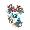
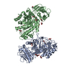
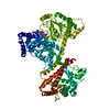
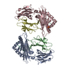
- PDB-3jux: Structure of the translocation ATPase SecA from Thermotoga maritima -
+ Open data

- Basic information
| Entry | Database: PDB / ID: 3jux | ||||||
|---|---|---|---|---|---|---|---|
| Title | Structure of the translocation ATPase SecA from Thermotoga maritima | ||||||
 Components Components | Protein translocase subunit secA | ||||||
 Keywords Keywords | PROTEIN TRANSPORT / protein translocation / ATPase / conformational change / peptide binding / ATP-binding / Cell inner membrane / Cell membrane / Cytoplasm / Membrane / Nucleotide-binding / Translocation / Transport | ||||||
| Function / homology |  Function and homology information Function and homology informationcell envelope Sec protein transport complex / protein-exporting ATPase activity / protein-secreting ATPase / protein transport by the Sec complex / intracellular protein transmembrane transport / protein import / protein targeting / ATP binding / plasma membrane / cytoplasm Similarity search - Function | ||||||
| Biological species |   Thermotoga maritima (bacteria) Thermotoga maritima (bacteria) | ||||||
| Method |  X-RAY DIFFRACTION / SYNCHROTRON / MOLECULAR REPLACEMENT / Resolution: 3.1 Å X-RAY DIFFRACTION / SYNCHROTRON / MOLECULAR REPLACEMENT / Resolution: 3.1 Å | ||||||
 Authors Authors | Zimmer, J. | ||||||
 Citation Citation | Journal: J.Mol.Biol. / Year: 2009 Title: Conformational flexibility and peptide interaction of the translocation ATPase SecA. Authors: Zimmer, J. / Rapoport, T.A. | ||||||
| History |
| ||||||
| Remark 650 | HELIX DETERMINATION METHOD: AUTHOR | ||||||
| Remark 700 | SHEET DETERMINATION METHOD: AUTHOR |
- Structure visualization
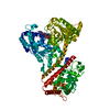
| Structure viewer | Molecule: MolmilJmol/JSmol |
|---|
- Downloads & links
-Download
| PDBx/mmCIF format | 3jux.cif.gz | 174.9 KB | Display | PDBx/mmCIF format |
|---|---|---|---|---|
| PDB format | pdb3jux.ent.gz | 136.7 KB | Display | PDB format |
| PDBx/mmJSON format | 3jux.json.gz | Tree view | PDBx/mmJSON format | |
| Others |  Other downloads Other downloads |
-Validation report
| Arichive directory | https://data.pdbj.org/pub/pdb/validation_reports/ju/3juxftp://data.pdbj.org/pub/pdb/validation_reports/ju/3jux | HTTPS FTP |
|---|
-Related structure data
| Related structure data | 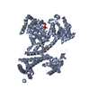 3jv2C 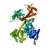 1tf5S S: Starting model for refinement C: citing same article ( |
|---|---|
| Similar structure data |


-Links
 PDBj
PDBj- Assembly
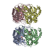
| Deposited unit | 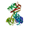
| ||||||||
|---|---|---|---|---|---|---|---|---|---|
| 1 |
| ||||||||
| Unit cell |
| ||||||||
| Details | monomer |
-Components
| #1: Protein | Mass: 94923.992 Da / Num. of mol.: 1 Source method: isolated from a genetically manipulated source Source: (gene. exp.) Thermotoga maritima (bacteria) / Strain: DSM 3109 / Gene: secA, TM_1578 / Plasmid: pET28a / Production host: |
|---|---|
| #2: Chemical | ChemComp-MG /   Mass: 24.305 Da / Num. of mol.: 1 / Source method: obtained synthetically / Formula: Mg Mass: 24.305 Da / Num. of mol.: 1 / Source method: obtained synthetically / Formula: Mg |
| #3: Chemical | ChemComp-ADP /   Mass: 427.201 Da / Num. of mol.: 1 / Source method: obtained synthetically / Formula: C10H15N5O10P2 / Comment: ADP, energy-carrying molecule*YM Mass: 427.201 Da / Num. of mol.: 1 / Source method: obtained synthetically / Formula: C10H15N5O10P2 / Comment: ADP, energy-carrying molecule*YM |
| #4: Water | ChemComp-HOH /  Mass: 18.015 Da / Num. of mol.: 2 / Source method: isolated from a natural source / Formula: H2O Mass: 18.015 Da / Num. of mol.: 2 / Source method: isolated from a natural source / Formula: H2O |
-Experimental details
-Experiment
| Experiment | Method: X-RAY DIFFRACTION / Number of used crystals: 3 |
|---|
- Sample preparation
| Crystal | Density Matthews: 2.45 Å3/Da / Density % sol: 49.83 % |
|---|---|
| Crystal grow | Temperature: 298 K / Method: vapor diffusion, sitting drop / pH: 7.5 Details: 0.1M Hepes pH 7.5, 60% MPD, VAPOR DIFFUSION, SITTING DROP, temperature 298K |
-Data collection
| Diffraction | Mean temperature: 100 K |
|---|---|
| Diffraction source | Source: SYNCHROTRON / Site: NSLS  / Beamline: X29A / Wavelength: 1.29 Å / Beamline: X29A / Wavelength: 1.29 Å |
| Detector | Type: ADSC QUANTUM 315 / Detector: CCD / Date: Feb 29, 2008 Details: double crystal monochromater with horizontal focusing sagittal bend second mono crystal with 4:1 magnification ratio and vertically focusing mirror. |
| Radiation | Monochromator: double crystal monochromater / Protocol: SINGLE WAVELENGTH / Monochromatic (M) / Laue (L): M / Scattering type: x-ray |
| Radiation wavelength | Wavelength: 1.29 Å / Relative weight: 1 |
| Reflection | Resolution: 3.1→50 Å / Num. all: 19431 / Num. obs: 17570 / % possible obs: 99.8 % / Observed criterion σ(F): 2 / Observed criterion σ(I): 1 / Redundancy: 6.8 % / Rsym value: 0.173 / Net I/σ(I): 13.6 |
| Reflection shell | Resolution: 3.1→3.22 Å / Redundancy: 6.7 % / Mean I/σ(I) obs: 2.5 / Num. unique all: 1913 / Rsym value: 0.79 / % possible all: 100 |
- Processing
| Software |
| |||||||||||||||||||||||||
|---|---|---|---|---|---|---|---|---|---|---|---|---|---|---|---|---|---|---|---|---|---|---|---|---|---|---|
| Refinement | Method to determine structure: MOLECULAR REPLACEMENT Starting model: 1TF5 Resolution: 3.1→30 Å / Isotropic thermal model: anisotropic / Cross valid method: THROUGHOUT / σ(F): 2 / σ(I): 1.5 / Stereochemistry target values: Engh & Huber
| |||||||||||||||||||||||||
| Displacement parameters | Biso mean: 42.3 Å2 | |||||||||||||||||||||||||
| Refinement step | Cycle: LAST / Resolution: 3.1→30 Å
| |||||||||||||||||||||||||
| LS refinement shell | Resolution: 3.1→3.13 Å /
|
