 Movie
Movie Controller


+ Open data

- Basic information
| Entry | Database: PDB / ID: 7apd | |||||||||
|---|---|---|---|---|---|---|---|---|---|---|
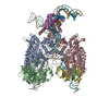
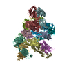
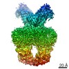
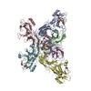
| Title | Bovine Papillomavirus E1 DNA helicase-replication fork complex | |||||||||
 Components Components |
| |||||||||
 Keywords Keywords | DNA BINDING PROTEIN / DNA / virus / helicase / replisome / DNA replication. | |||||||||
| Function / homology |  Function and homology information Function and homology information3'-5' DNA helicase activity / DNA 3'-5' helicase / DNA replication / host cell nucleus / ATP hydrolysis activity / DNA binding / ATP binding Similarity search - Function | |||||||||
| Biological species |  Bovine papillomavirus Bovine papillomavirus | |||||||||
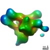
| Method | ELECTRON MICROSCOPY / single particle reconstruction / cryo EM / Resolution: 3.9 Å | |||||||||
 Authors Authors | Javed, A. / Major, B. / Stead, J. / Sanders, C.M. / Orlova, E.V. | |||||||||
| Funding support |  United Kingdom, 2items United Kingdom, 2items
| |||||||||
 Citation Citation | Journal: Nat Commun / Year: 2021 Title: Unwinding of a DNA replication fork by a hexameric viral helicase. Authors: Abid Javed / Balazs Major / Jonathan A Stead / Cyril M Sanders / Elena V Orlova / Abstract: Hexameric helicases are motor proteins that unwind double-stranded DNA (dsDNA) during DNA replication but how they are optimised for strand separation is unclear. Here we present the cryo-EM ...Hexameric helicases are motor proteins that unwind double-stranded DNA (dsDNA) during DNA replication but how they are optimised for strand separation is unclear. Here we present the cryo-EM structure of the full-length E1 helicase from papillomavirus, revealing all arms of a bound DNA replication fork and their interactions with the helicase. The replication fork junction is located at the entrance to the helicase collar ring, that sits above the AAA + motor assembly. dsDNA is escorted to and the 5´ single-stranded DNA (ssDNA) away from the unwinding point by the E1 dsDNA origin binding domains. The 3´ ssDNA interacts with six spirally-arranged β-hairpins and their cyclical top-to-bottom movement pulls the ssDNA through the helicase. Pulling of the RF against the collar ring separates the base-pairs, while modelling of the conformational cycle suggest an accompanying movement of the collar ring has an auxiliary role, helping to make efficient use of ATP in duplex unwinding. | |||||||||
| History |
|
- Structure visualization
| Movie |
Movie viewer |
|---|---|
| Structure viewer | Molecule: MolmilJmol/JSmol |
- Downloads & links
-Download
| PDBx/mmCIF format | 7apd.cif.gz | 380.8 KB | Display | PDBx/mmCIF format |
|---|---|---|---|---|
| PDB format | pdb7apd.ent.gz | 297.3 KB | Display | PDB format |
| PDBx/mmJSON format | 7apd.json.gz | Tree view | PDBx/mmJSON format | |
| Others |  Other downloads Other downloads |
-Validation report
| Arichive directory | https://data.pdbj.org/pub/pdb/validation_reports/ap/7apdftp://data.pdbj.org/pub/pdb/validation_reports/ap/7apd | HTTPS FTP |
|---|
-Related structure data
| Related structure data |  11852MC M: map data used to model this data C: citing same article ( |
|---|---|
| Similar structure data |


-Links
 PDBj
PDBj


- Assembly
| Deposited unit | 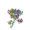
|
|---|---|
| 1 |
|
-Components
| #1: Protein | Mass: 17162.084 Da / Num. of mol.: 2 Source method: isolated from a genetically manipulated source Details: OBD domains from subunits B and E. / Source: (gene. exp.) Bovine papillomavirus / Gene: E1 / Production host:  #2: Protein | Mass: 33859.172 Da / Num. of mol.: 6 Source method: isolated from a genetically manipulated source Source: (gene. exp.) Bovine papillomavirus / Gene: E1 / Production host: #3: DNA chain | | Mass: 12142.779 Da / Num. of mol.: 1 / Source method: obtained synthetically / Details: 5'-3' ssDNA strand of the DNA replication fork. / Source: (synth.) Bovine papillomavirus#4: DNA chain | | Mass: 11080.090 Da / Num. of mol.: 1 / Source method: obtained synthetically / Details: 3'-5' ssDNA strand of the DNA replication fork. / Source: (synth.) Bovine papillomavirus |
|---|
-Experimental details
-Experiment
| Experiment | Method: ELECTRON MICROSCOPY |
|---|---|
| EM experiment | Aggregation state: PARTICLE / 3D reconstruction method: single particle reconstruction |
- Sample preparation
| Component |
| ||||||||||||||||||||||||||||
|---|---|---|---|---|---|---|---|---|---|---|---|---|---|---|---|---|---|---|---|---|---|---|---|---|---|---|---|---|---|
| Molecular weight | Value: 0.4134 MDa / Experimental value: YES | ||||||||||||||||||||||||||||
| Source (natural) |
| ||||||||||||||||||||||||||||
| Source (recombinant) |
| ||||||||||||||||||||||||||||
| Buffer solution | pH: 7.2 | ||||||||||||||||||||||||||||
| Buffer component |
| ||||||||||||||||||||||||||||
| Specimen | Conc.: 0.05 mg/ml / Embedding applied: NO / Shadowing applied: NO / Staining applied: NO / Vitrification applied: YES | ||||||||||||||||||||||||||||
| Specimen support | Grid material: COPPER / Grid type: PELCO Ultrathin Carbon with Lacey Carbon | ||||||||||||||||||||||||||||
| Vitrification | Instrument: FEI VITROBOT MARK IV / Cryogen name: ETHANE / Humidity: 100 % / Chamber temperature: 281 K Details: 3 ul of sample was applied Lacey ultra-thin carbon film grids. |
- Electron microscopy imaging
| Experimental equipment |  Model: Titan Krios / Image courtesy: FEI Company |
|---|---|
| Microscopy | Model: FEI TITAN KRIOS |
| Electron gun | Electron source:  FIELD EMISSION GUN / Accelerating voltage: 300 kV / Illumination mode: FLOOD BEAM FIELD EMISSION GUN / Accelerating voltage: 300 kV / Illumination mode: FLOOD BEAM |
| Electron lens | Mode: BRIGHT FIELD / Nominal magnification: 81000 X / Calibrated magnification: 47170 X / Nominal defocus max: 2500 nm / Nominal defocus min: 1200 nm / Cs: 2.7 mm / C2 aperture diameter: 70 µm / Alignment procedure: ZEMLIN TABLEAU |
| Specimen holder | Cryogen: NITROGEN / Specimen holder model: FEI TITAN KRIOS AUTOGRID HOLDER / Temperature (max): 98 K / Temperature (min): 95 K |
| Image recording | Average exposure time: 3 sec. / Electron dose: 50.4 e/Å2 / Film or detector model: GATAN K3 BIOQUANTUM (6k x 4k) / Num. of grids imaged: 2 / Num. of real images: 12136 |
| EM imaging optics | Energyfilter name: GIF Quantum LS / Energyfilter slit width: 20 eV |
| Image scans | Sampling size: 5.2 µm / Width: 5760 / Height: 4092 |
- Processing
| EM software |
| |||||||||||||||||||||||||||||||||||||||||||||||||||||||||||||||||
|---|---|---|---|---|---|---|---|---|---|---|---|---|---|---|---|---|---|---|---|---|---|---|---|---|---|---|---|---|---|---|---|---|---|---|---|---|---|---|---|---|---|---|---|---|---|---|---|---|---|---|---|---|---|---|---|---|---|---|---|---|---|---|---|---|---|---|
| CTF correction | Type: PHASE FLIPPING AND AMPLITUDE CORRECTION | |||||||||||||||||||||||||||||||||||||||||||||||||||||||||||||||||
| Particle selection | Num. of particles selected: 568120 | |||||||||||||||||||||||||||||||||||||||||||||||||||||||||||||||||
| Symmetry | Point symmetry: C1 (asymmetric) | |||||||||||||||||||||||||||||||||||||||||||||||||||||||||||||||||
| 3D reconstruction | Resolution: 3.9 Å / Resolution method: FSC 0.143 CUT-OFF / Num. of particles: 81831 / Algorithm: FOURIER SPACE / Num. of class averages: 1 / Symmetry type: POINT | |||||||||||||||||||||||||||||||||||||||||||||||||||||||||||||||||
| Atomic model building | B value: 102 / Protocol: FLEXIBLE FIT / Space: REAL / Target criteria: Correlation coefficient | |||||||||||||||||||||||||||||||||||||||||||||||||||||||||||||||||
| Atomic model building |
|


