[English] 日本語
Yorodumi
- PDB-2gxa: Crystal structure of papillomavirus E1 hexameric helicase with ss... -

+
Open data


ID or keywords:

Loading...

-
Basic information

Entry
Database: PDB / ID: 2gxa
Title
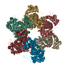
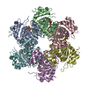
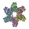
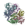
Crystal structure of papillomavirus E1 hexameric helicase with ssDNA and MgADP
Components
  • 5'-D(*TP*TP*TP*TP*TP*TP*TP*TP*TP*TP*TP*TP*T)-3'
  • Replication protein E1
Keywordsreplication/DNA / DNA helicase / AAA+ / ATPase / replication / virus / initiator protein / replication-DNA COMPLEX
Function / homology
Function and homology information


DNA helicase / forked DNA-dependent helicase activity / single-stranded 3'-5' DNA helicase activity / four-way junction helicase activity / double-stranded DNA helicase activity / DNA replication / host cell nucleus / ATP hydrolysis activity / DNA binding / ATP binding
Similarity search - Function
Zinc finger, large T-antigen D1 domain / DNA helicase E1, C-terminal, Papillomavirus / DNA helicase E1, N-terminal, Papillomavirus / Replication protein E1, papillomavirus / DNA helicase E1, DNA-binding domain, papillomavirus / DNA helicase E1, DNA-binding domain superfamily, papillomavirus / Papillomavirus helicase / E1 Protein, N terminal domain / Papillomavirus E1, DNA-binding domain / Zinc finger, large T-antigen D1 domain superfamily ...Zinc finger, large T-antigen D1 domain / DNA helicase E1, C-terminal, Papillomavirus / DNA helicase E1, N-terminal, Papillomavirus / Replication protein E1, papillomavirus / DNA helicase E1, DNA-binding domain, papillomavirus / DNA helicase E1, DNA-binding domain superfamily, papillomavirus / Papillomavirus helicase / E1 Protein, N terminal domain / Papillomavirus E1, DNA-binding domain / Zinc finger, large T-antigen D1 domain superfamily / Helicase, superfamily 3, DNA virus / Superfamily 3 helicase of DNA viruses domain profile. / Arc Repressor Mutant, subunit A / P-loop containing nucleotide triphosphate hydrolases / Rossmann fold / P-loop containing nucleoside triphosphate hydrolase / Orthogonal Bundle / 3-Layer(aba) Sandwich / Mainly Alpha / Alpha Beta
Similarity search - Domain/homology
ADENOSINE-5'-DIPHOSPHATE / DNA / DNA (> 10) / Replication protein E1
Similarity search - Component
Biological speciesBovine papillomavirus type 1
MethodX-RAY DIFFRACTION / SYNCHROTRON / MOLECULAR REPLACEMENT / Resolution: 3.15 Å
AuthorsEnemark, E.J. / Joshua-Tor, L.
CitationJournal: Nature / Year: 2006
Title: Mechanism of DNA translocation in a replicative hexameric helicase.
Authors: Enemark, E.J. / Joshua-Tor, L.
History
DepositionMay 8, 2006Deposition site: RCSB / Processing site: RCSB
Revision 1.0Jul 25, 2006Provider: repository / Type: Initial release
Revision 1.1May 1, 2008Group: Version format compliance
Revision 1.2Jul 13, 2011Group: Version format compliance
Revision 1.3Aug 30, 2023Group: Data collection / Database references ...Data collection / Database references / Derived calculations / Refinement description
Category: chem_comp_atom / chem_comp_bond ...chem_comp_atom / chem_comp_bond / database_2 / pdbx_initial_refinement_model / pdbx_struct_conn_angle / struct_conn / struct_site
Item: _database_2.pdbx_DOI / _database_2.pdbx_database_accession ..._database_2.pdbx_DOI / _database_2.pdbx_database_accession / _pdbx_struct_conn_angle.ptnr1_auth_asym_id / _pdbx_struct_conn_angle.ptnr1_auth_comp_id / _pdbx_struct_conn_angle.ptnr1_auth_seq_id / _pdbx_struct_conn_angle.ptnr1_label_asym_id / _pdbx_struct_conn_angle.ptnr1_label_atom_id / _pdbx_struct_conn_angle.ptnr1_label_comp_id / _pdbx_struct_conn_angle.ptnr1_label_seq_id / _pdbx_struct_conn_angle.ptnr3_auth_asym_id / _pdbx_struct_conn_angle.ptnr3_auth_comp_id / _pdbx_struct_conn_angle.ptnr3_auth_seq_id / _pdbx_struct_conn_angle.ptnr3_label_asym_id / _pdbx_struct_conn_angle.ptnr3_label_atom_id / _pdbx_struct_conn_angle.ptnr3_label_comp_id / _pdbx_struct_conn_angle.ptnr3_label_seq_id / _pdbx_struct_conn_angle.value / _struct_conn.pdbx_dist_value / _struct_conn.ptnr1_auth_comp_id / _struct_conn.ptnr1_auth_seq_id / _struct_conn.ptnr1_label_asym_id / _struct_conn.ptnr1_label_atom_id / _struct_conn.ptnr1_label_comp_id / _struct_conn.ptnr2_auth_asym_id / _struct_conn.ptnr2_auth_comp_id / _struct_conn.ptnr2_auth_seq_id / _struct_conn.ptnr2_label_asym_id / _struct_conn.ptnr2_label_atom_id / _struct_conn.ptnr2_label_comp_id / _struct_conn.ptnr2_label_seq_id / _struct_site.pdbx_auth_asym_id / _struct_site.pdbx_auth_comp_id / _struct_site.pdbx_auth_seq_id

-
Structure visualization

Structure viewerMolecule:
MolmilJmol/JSmol

Downloads & links

-
Assembly

Deposited unit
M: 5'-D(*TP*TP*TP*TP*TP*TP*TP*TP*TP*TP*TP*TP*T)-3'
N: 5'-D(*TP*TP*TP*TP*TP*TP*TP*TP*TP*TP*TP*TP*T)-3'
A: Replication protein E1
B: Replication protein E1
C: Replication protein E1
D: Replication protein E1
E: Replication protein E1
F: Replication protein E1
G: Replication protein E1
H: Replication protein E1
I: Replication protein E1
J: Replication protein E1
K: Replication protein E1
L: Replication protein E1
hetero molecules


Theoretical massNumber of molelcules
Total (without water)386,34938
Polymers381,30014
Non-polymers5,04924
Water27015
1
M: 5'-D(*TP*TP*TP*TP*TP*TP*TP*TP*TP*TP*TP*TP*T)-3'
A: Replication protein E1
B: Replication protein E1
C: Replication protein E1
D: Replication protein E1
E: Replication protein E1
F: Replication protein E1
hetero molecules


Theoretical massNumber of molelcules
Total (without water)193,40620
Polymers190,6507
Non-polymers2,75613
Water724
TypeNameSymmetry operationNumber
identity operation1_555x,y,z1
2
N: 5'-D(*TP*TP*TP*TP*TP*TP*TP*TP*TP*TP*TP*TP*T)-3'
G: Replication protein E1
H: Replication protein E1
I: Replication protein E1
J: Replication protein E1
K: Replication protein E1
L: Replication protein E1
hetero molecules


Theoretical massNumber of molelcules
Total (without water)192,94318
Polymers190,6507
Non-polymers2,29311
Water543
TypeNameSymmetry operationNumber
identity operation1_555x,y,z1
Unit cell
Length a, b, c (Å)100.510, 100.881, 125.023
Angle α, β, γ (deg.)92.60, 111.46, 106.01
Int Tables number1
Space group name H-MP1
Details
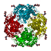
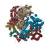
The structure consists of 2 independent hexamers in complex with ssDNA and MgADP. Each is a unique biological assembly (hexamer 1: chains A-F, M, O1-6, X1-6; hexamer 2: chains G-L, N, O7-11, X7-11 ).

-
Components

-
DNA chain / Protein , 2 types, 14 molecules MNABCDEFGHIJKL

#1: DNA chain 5'-D(*TP*TP*TP*TP*TP*TP*TP*TP*TP*TP*TP*TP*T)-3'


Mass: 3909.549 Da / Num. of mol.: 2 / Source method: obtained synthetically
#2: Protein
Replication protein E1


Mass: 31123.447 Da / Num. of mol.: 12
Source method: isolated from a genetically manipulated source
Source: (gene. exp.) Bovine papillomavirus type 1 / Genus: Deltapapillomavirus / Species: Bovine papillomavirus - 1 / Strain: BPV-1 / Gene: E1 / Plasmid: pGEX-4T-1 / Species (production host): Escherichia coli / Production host: Escherichia coli BL21(DE3) (bacteria) / Strain (production host): BL21(DE3) / References: UniProt: P03116

-
Non-polymers , 4 types, 39 molecules

#3: Chemical
ChemComp-MG / MAGNESIUM ION


Mass: 24.305 Da / Num. of mol.: 10 / Source method: obtained synthetically / Formula: Mg
#4: Chemical ChemComp-CL / CHLORIDE ION


Mass: 35.453 Da / Num. of mol.: 3 / Source method: obtained synthetically / Formula: Cl
#5: Chemical
ChemComp-ADP / ADENOSINE-5'-DIPHOSPHATE


Mass: 427.201 Da / Num. of mol.: 11 / Source method: obtained synthetically / Formula: C10H15N5O10P2 / Comment: ADP, energy-carrying molecule*YM
#6: Water ChemComp-HOH / water


Mass: 18.015 Da / Num. of mol.: 15 / Source method: isolated from a natural source / Formula: H2O

-
Experimental details

-
Experiment

ExperimentMethod: X-RAY DIFFRACTION / Number of used crystals: 1

-
Sample preparation

CrystalDensity Matthews: 2.67 Å3/Da / Density % sol: 53.95 %
Crystal growTemperature: 290 K / Method: vapor diffusion, hanging drop / pH: 7.5
Details: 100 mM KCl, pH 7.5, VAPOR DIFFUSION, HANGING DROP, temperature 290K
Components of the solutions
IDNameCrystal-IDSol-ID
1KCl11
2H2O11
3KCL12
4H2O12

-
Data collection

DiffractionMean temperature: 100 K
Diffraction sourceSource: SYNCHROTRON / Site: NSLS / Beamline: X29A / Wavelength: 1.1 Å
DetectorType: ADSC QUANTUM 315 / Detector: CCD
RadiationProtocol: SINGLE WAVELENGTH / Monochromatic (M) / Laue (L): M / Scattering type: x-ray
Radiation wavelengthWavelength: 1.1 Å / Relative weight: 1
ReflectionResolution: 3.15→50 Å / Num. all: 75701 / Num. obs: 74589 / % possible obs: 98.6 % / Observed criterion σ(I): -3 / Redundancy: 3.7 % / Biso Wilson estimate: 55.1 Å2 / Rmerge(I) obs: 0.1 / Net I/σ(I): 10.5
Reflection shellResolution: 3.15→3.26 Å / Redundancy: 3.7 % / Rmerge(I) obs: 0.718 / Mean I/σ(I) obs: 1.2 / Num. unique all: 7550 / % possible all: 98.1

-
Processing

Software
NameVersionClassification
CNS1.1refinement
CBASSdata collection
HKL-2000data scaling
PHASERphasing
RefinementMethod to determine structure: MOLECULAR REPLACEMENT
Starting model: composite of PDB ENTRY 1TUE and unpublished structure
Resolution: 3.15→44.65 Å / Rfactor Rfree error: 0.005 / Data cutoff high absF: 41133.91 / Data cutoff low absF: 0 / Isotropic thermal model: GROUP / Cross valid method: THROUGHOUT / σ(F): 0 / Stereochemistry target values: Engh & Huber
RfactorNum. reflection% reflectionSelection details
Rfree0.298 3626 5.1 %RANDOM
Rwork0.239 ---
obs0.239 71517 91.9 %-
all-71517 --
Solvent computationSolvent model: FLAT MODEL / Bsol: 39.9217 Å2 / ksol: 0.246589 e/Å3
Displacement parametersBiso mean: 74.6 Å2
Baniso -1Baniso -2Baniso -3
1-3.21 Å25.07 Å214.02 Å2
2---10.63 Å2-2.67 Å2
3---7.42 Å2
Refine analyze
FreeObs
Luzzati coordinate error0.53 Å0.4 Å
Luzzati d res low-5 Å
Luzzati sigma a0.73 Å0.65 Å
Refinement stepCycle: LAST / Resolution: 3.15→44.65 Å
ProteinNucleic acidLigandSolventTotal
Num. atoms25787 262 310 15 26374
Refine LS restraints
Refine-IDTypeDev ideal
X-RAY DIFFRACTIONc_bond_d0.009
X-RAY DIFFRACTIONc_angle_deg1.4
X-RAY DIFFRACTIONc_dihedral_angle_d21.5
X-RAY DIFFRACTIONc_improper_angle_d0.89
LS refinement shellResolution: 3.15→3.29 Å / Rfactor Rfree error: 0.019 / Total num. of bins used: 6
RfactorNum. reflection% reflection
Rfree0.431 493 5.3 %
Rwork0.388 8802 -
obs--70.9 %
Xplor file
Refine-IDSerial noParam fileTopol file
X-RAY DIFFRACTION1protein_rep.paramprotein.top
X-RAY DIFFRACTION2ion.paramion.top
X-RAY DIFFRACTION3water.paramwater.top
X-RAY DIFFRACTION4adp.paramadp.top
X-RAY DIFFRACTION5dna-rna_rep.paramdna-rna.top

+
About Yorodumi

-
News

-
Feb 9, 2022. New format data for meta-information of EMDB entries

New format data for meta-information of EMDB entries

  • Version 3 of the EMDB header file is now the official format.
  • The previous official version 1.9 will be removed from the archive.

Related info.:EMDB header

External links:wwPDB to switch to version 3 of the EMDB data model

-
Aug 12, 2020. Covid-19 info

Covid-19 info

URL: https://pdbj.org/emnavi/covid19.php

New page: Covid-19 featured information page in EM Navigator.

Related info.:Covid-19 info / Mar 5, 2020. Novel coronavirus structure data

+
Mar 5, 2020. Novel coronavirus structure data

Novel coronavirus structure data

Related info.:Yorodumi Speices / Aug 12, 2020. Covid-19 info

External links:COVID-19 featured content - PDBj / Molecule of the Month (242):Coronavirus Proteases

+
Jan 31, 2019. EMDB accession codes are about to change! (news from PDBe EMDB page)

EMDB accession codes are about to change! (news from PDBe EMDB page)

  • The allocation of 4 digits for EMDB accession codes will soon come to an end. Whilst these codes will remain in use, new EMDB accession codes will include an additional digit and will expand incrementally as the available range of codes is exhausted. The current 4-digit format prefixed with “EMD-” (i.e. EMD-XXXX) will advance to a 5-digit format (i.e. EMD-XXXXX), and so on. It is currently estimated that the 4-digit codes will be depleted around Spring 2019, at which point the 5-digit format will come into force.
  • The EM Navigator/Yorodumi systems omit the EMD- prefix.

Related info.:Q: What is EMD? / ID/Accession-code notation in Yorodumi/EM Navigator

External links:EMDB Accession Codes are Changing Soon! / Contact to PDBj

+
Jul 12, 2017. Major update of PDB

Major update of PDB

  • wwPDB released updated PDB data conforming to the new PDBx/mmCIF dictionary.
  • This is a major update changing the version number from 4 to 5, and with Remediation, in which all the entries are updated.
  • In this update, many items about electron microscopy experimental information are reorganized (e.g. em_software).
  • Now, EM Navigator and Yorodumi are based on the updated data.

External links:wwPDB Remediation / Enriched Model Files Conforming to OneDep Data Standards Now Available in the PDB FTP Archive

-
Yorodumi

Thousand views of thousand structures

  • Yorodumi is a browser for structure data from EMDB, PDB, SASBDB, etc.
  • This page is also the successor to EM Navigator detail page, and also detail information page/front-end page for Omokage search.
  • The word "yorodu" (or yorozu) is an old Japanese word meaning "ten thousand". "mi" (miru) is to see.

Related info.:EMDB / PDB / SASBDB / Comparison of 3 databanks / Yorodumi Search / Aug 31, 2016. New EM Navigator & Yorodumi / Yorodumi Papers / Jmol/JSmol / Function and homology information / Changes in new EM Navigator and Yorodumi

Read more