 Movie
Movie Controller


+ Open data

- Basic information
| Entry | Database: PDB / ID: 7aog | |||||||||
|---|---|---|---|---|---|---|---|---|---|---|
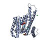
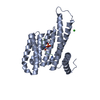
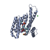
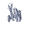
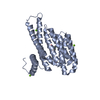
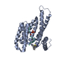
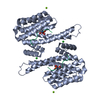
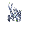
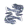
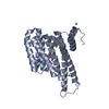
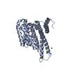
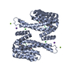
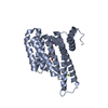
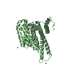
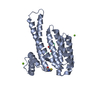
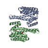
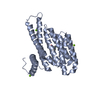
| Title | 14-3-3 sigma in complex with Pin1 binding site pS72 | |||||||||
 Components Components |
| |||||||||
 Keywords Keywords | PEPTIDE BINDING PROTEIN / 1433 / Peptidyl-prolyl cis-trans isomerase NIMA-interacting 1 | |||||||||
| Function / homology |  Function and homology information Function and homology informationcis-trans isomerase activity / phosphothreonine residue binding / negative regulation of cell motility / negative regulation of brown fat cell differentiation / regulation of protein localization to nucleus / mitogen-activated protein kinase kinase binding / ubiquitin ligase activator activity / GTPase activating protein binding / : / protein peptidyl-prolyl isomerization ...cis-trans isomerase activity / phosphothreonine residue binding / negative regulation of cell motility / negative regulation of brown fat cell differentiation / regulation of protein localization to nucleus / mitogen-activated protein kinase kinase binding / ubiquitin ligase activator activity / GTPase activating protein binding / : / protein peptidyl-prolyl isomerization / regulation of mitotic nuclear division / regulation of epidermal cell division / protein kinase C inhibitor activity / positive regulation of epidermal cell differentiation / keratinocyte development / keratinization / negative regulation of SMAD protein signal transduction / PI5P Regulates TP53 Acetylation / regulation of cell-cell adhesion / negative regulation of amyloid-beta formation / establishment of skin barrier / cytoskeletal motor activity / Regulation of localization of FOXO transcription factors / keratinocyte proliferation / negative regulation of keratinocyte proliferation / phosphoserine residue binding / Activation of BAD and translocation to mitochondria / RHO GTPases Activate NADPH Oxidases / cAMP/PKA signal transduction / negative regulation of protein localization to plasma membrane / SARS-CoV-2 targets host intracellular signalling and regulatory pathways / postsynaptic cytosol / negative regulation of stem cell proliferation / negative regulation of protein kinase activity / Rho protein signal transduction / SARS-CoV-1 targets host intracellular signalling and regulatory pathways / RHO GTPases activate PKNs / Chk1/Chk2(Cds1) mediated inactivation of Cyclin B:Cdk1 complex / positive regulation of protein localization / protein export from nucleus / TP53 Regulates Transcription of Genes Involved in G2 Cell Cycle Arrest / negative regulation of innate immune response / positive regulation of cell adhesion / release of cytochrome c from mitochondria / positive regulation of protein export from nucleus / stem cell proliferation / regulation of cytokinesis / peptidylprolyl isomerase / TP53 Regulates Metabolic Genes / peptidyl-prolyl cis-trans isomerase activity / Translocation of SLC2A4 (GLUT4) to the plasma membrane / protein sequestering activity / Negative regulators of DDX58/IFIH1 signaling / negative regulation of transforming growth factor beta receptor signaling pathway / regulation of protein stability / phosphoprotein binding / negative regulation of protein catabolic process / negative regulation of ERK1 and ERK2 cascade / positive regulation of protein phosphorylation / synapse organization / beta-catenin binding / protein destabilization / tau protein binding / ISG15 antiviral mechanism / intrinsic apoptotic signaling pathway in response to DNA damage / neuron differentiation / intracellular protein localization / positive regulation of canonical Wnt signaling pathway / regulation of protein localization / positive regulation of cell growth / regulation of gene expression / sperm midpiece / midbody / cellular response to hypoxia / Regulation of TP53 Activity through Phosphorylation / response to hypoxia / regulation of cell cycle / nuclear speck / protein stabilization / ciliary basal body / cadherin binding / protein kinase binding / glutamatergic synapse / negative regulation of transcription by RNA polymerase II / signal transduction / positive regulation of transcription by RNA polymerase II / : / extracellular exosome / nucleoplasm / identical protein binding / nucleus / cytoplasm / cytosol Similarity search - Function | |||||||||
| Biological species |  Homo sapiens (human) Homo sapiens (human) | |||||||||
| Method |  X-RAY DIFFRACTION / SYNCHROTRON / MOLECULAR REPLACEMENT / molecular replacement / Resolution: 1.5 Å X-RAY DIFFRACTION / SYNCHROTRON / MOLECULAR REPLACEMENT / molecular replacement / Resolution: 1.5 Å | |||||||||
 Authors Authors | Wolter, M. / Dijck, L.v. / Cossar, P.J. / Ottmann, C. | |||||||||
| Funding support | 2items
| |||||||||
 Citation Citation | Journal: J.Am.Chem.Soc. / Year: 2021 Title: Reversible Covalent Imine-Tethering for Selective Stabilization of 14-3-3 Hub Protein Interactions. Authors: Cossar, P.J. / Wolter, M. / van Dijck, L. / Valenti, D. / Levy, L.M. / Ottmann, C. / Brunsveld, L. | |||||||||
| History |
|
- Structure visualization
| Structure viewer | Molecule: MolmilJmol/JSmol |
|---|
- Downloads & links
-Download
| PDBx/mmCIF format | 7aog.cif.gz | 112.5 KB | Display | PDBx/mmCIF format |
|---|---|---|---|---|
| PDB format | pdb7aog.ent.gz | 85.5 KB | Display | PDB format |
| PDBx/mmJSON format | 7aog.json.gz | Tree view | PDBx/mmJSON format | |
| Others |  Other downloads Other downloads |
-Validation report
| Arichive directory | https://data.pdbj.org/pub/pdb/validation_reports/ao/7aogftp://data.pdbj.org/pub/pdb/validation_reports/ao/7aog | HTTPS FTP |
|---|
-Related structure data
| Related structure data | 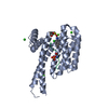 7axnC 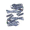 7ayfC 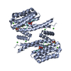 7az1C 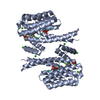 7az2C  7bdpC  7bdtC  7bdyC  7bfwC 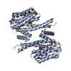 7bg3C 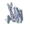 7bgqC 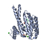 7bgrC 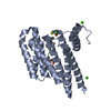 7bgvC  7bgwC  7nifC  7nigC  7nixC 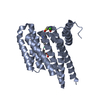 7nj6C 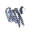 7nj8C 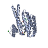 7njaC 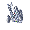 7nqpC 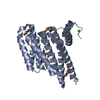 7nrkC 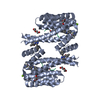 7nrlC 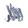 7nsvC 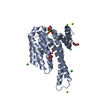 3mhrS S: Starting model for refinement C: citing same article ( |
|---|---|
| Similar structure data |


-Links
 PDBj
PDBj


- Assembly
| Deposited unit | 
| ||||||||
|---|---|---|---|---|---|---|---|---|---|
| 1 | 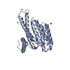
| ||||||||
| Unit cell |
| ||||||||
| Components on special symmetry positions |
|
-Components
| #1: Protein | Mass: 28226.518 Da / Num. of mol.: 1 Source method: isolated from a genetically manipulated source Source: (gene. exp.) Homo sapiens (human) / Gene: SFN, HME1 / Production host:  | ||||||||
|---|---|---|---|---|---|---|---|---|---|
| #2: Protein/peptide | Mass: 2195.378 Da / Num. of mol.: 1 / Source method: obtained synthetically / Source: (synth.) Homo sapiens (human) / References: UniProt: Q13526, peptidylprolyl isomerase | ||||||||
| #3: Chemical |   Mass: 40.078 Da / Num. of mol.: 2 / Source method: obtained synthetically / Formula: Ca Mass: 40.078 Da / Num. of mol.: 2 / Source method: obtained synthetically / Formula: Ca#4: Chemical |   Mass: 35.453 Da / Num. of mol.: 2 / Source method: obtained synthetically / Formula: Cl Mass: 35.453 Da / Num. of mol.: 2 / Source method: obtained synthetically / Formula: Cl#5: Water | ChemComp-HOH / |  Mass: 18.015 Da / Num. of mol.: 284 / Source method: isolated from a natural source / Formula: H2O Mass: 18.015 Da / Num. of mol.: 284 / Source method: isolated from a natural source / Formula: H2OHas ligand of interest | N | Has protein modification | Y | |
-Experimental details
-Experiment
| Experiment | Method: X-RAY DIFFRACTION / Number of used crystals: 1 |
|---|
- Sample preparation
| Crystal | Density Matthews: 2.38 Å3/Da / Density % sol: 48.3 % |
|---|---|
| Crystal grow | Temperature: 277 K / Method: vapor diffusion, hanging drop Details: 0.095 M HEPES Na pH 7.1, 27% PEG400, 0.19M Calcium chloride, 5% Glycerol |
-Data collection
| Diffraction | Mean temperature: 100 K / Serial crystal experiment: N |
|---|---|
| Diffraction source | Source: SYNCHROTRON / Site: PETRA III, DESY  / Beamline: P11 / Wavelength: 1.0332 Å / Beamline: P11 / Wavelength: 1.0332 Å |
| Detector | Type: DECTRIS PILATUS 6M-F / Detector: PIXEL / Date: Jun 22, 2019 |
| Radiation | Protocol: SINGLE WAVELENGTH / Monochromatic (M) / Laue (L): M / Scattering type: x-ray |
| Radiation wavelength | Wavelength: 1.0332 Å / Relative weight: 1 |
| Reflection | Resolution: 1.5→62.65 Å / Num. obs: 44552 / % possible obs: 95.1 % / Redundancy: 10.5 % / Biso Wilson estimate: 19.38 Å2 / CC1/2: 0.999 / Rmerge(I) obs: 0.055 / Rpim(I) all: 0.017 / Rrim(I) all: 0.058 / Net I/σ(I): 20.1 |
| Reflection shell | Resolution: 1.5→1.53 Å / Redundancy: 3.8 % / Rmerge(I) obs: 0.736 / Mean I/σ(I) obs: 1.6 / Num. unique obs: 1508 / CC1/2: 0.527 / Rpim(I) all: 0.425 / Rrim(I) all: 0.859 / % possible all: 64.3 |
-Phasing
| Phasing | Method: molecular replacement |
|---|
- Processing
| Software |
| ||||||||||||||||||||||||||||||||||||||||||||||||||||||||||||||||||||||||||||||||||||||||||||||||
|---|---|---|---|---|---|---|---|---|---|---|---|---|---|---|---|---|---|---|---|---|---|---|---|---|---|---|---|---|---|---|---|---|---|---|---|---|---|---|---|---|---|---|---|---|---|---|---|---|---|---|---|---|---|---|---|---|---|---|---|---|---|---|---|---|---|---|---|---|---|---|---|---|---|---|---|---|---|---|---|---|---|---|---|---|---|---|---|---|---|---|---|---|---|---|---|---|---|
| Refinement | Method to determine structure: MOLECULAR REPLACEMENT Starting model: 3MHR Resolution: 1.5→56.029 Å / SU ML: 0.15 / Cross valid method: FREE R-VALUE / σ(F): 1.35 / Phase error: 18.86 / Stereochemistry target values: ML
| ||||||||||||||||||||||||||||||||||||||||||||||||||||||||||||||||||||||||||||||||||||||||||||||||
| Solvent computation | Shrinkage radii: 0.9 Å / VDW probe radii: 1.11 Å / Solvent model: FLAT BULK SOLVENT MODEL | ||||||||||||||||||||||||||||||||||||||||||||||||||||||||||||||||||||||||||||||||||||||||||||||||
| Displacement parameters | Biso max: 91.04 Å2 / Biso mean: 26.3518 Å2 / Biso min: 12.97 Å2 | ||||||||||||||||||||||||||||||||||||||||||||||||||||||||||||||||||||||||||||||||||||||||||||||||
| Refinement step | Cycle: final / Resolution: 1.5→56.029 Å
| ||||||||||||||||||||||||||||||||||||||||||||||||||||||||||||||||||||||||||||||||||||||||||||||||
| Refine LS restraints |
| ||||||||||||||||||||||||||||||||||||||||||||||||||||||||||||||||||||||||||||||||||||||||||||||||
| LS refinement shell | Refine-ID: X-RAY DIFFRACTION / Rfactor Rfree error: 0
|
